Volume 20, Number 12—December 2014
Dispatch
Equine Influenza A(H3N8) Virus Infection in Cats
Shuo Su1, Lifang Wang1, Xinliang Fu, Shuyi He, Malin Hong, Pei Zhou, Alexander Lai , Gregory C. Gray, and Shoujun Li
Figure 2
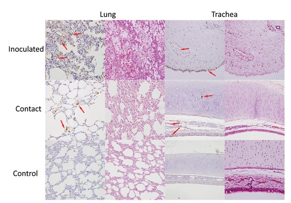
Figure 2. Detection of viral antigens in the respiratory tract of cats inoculated with equine influenza A(H3N8) virus and from a contact cohort. For each tissue type, the left column shows incubation with a monoclonal antibody against equine influenza virus hemagglutinin and the right column shows hematoxylin and eosin staining. Arrows indicate detection of viral antigen (hemagglutinin) expression (brownish staining). Original magnification ×100.
1These authors contributed equally to this work.
Page created: November 19, 2014
Page updated: November 19, 2014
Page reviewed: November 19, 2014
The conclusions, findings, and opinions expressed by authors contributing to this journal do not necessarily reflect the official position of the U.S. Department of Health and Human Services, the Public Health Service, the Centers for Disease Control and Prevention, or the authors' affiliated institutions. Use of trade names is for identification only and does not imply endorsement by any of the groups named above.