Volume 21, Number 2—February 2015
Online Report
Melioidosis Diagnostic Workshop, 20131
Culture and Identification
Rapid Detection of B. pseudomallei in Clinical Specimens
Serologic Tests
Future Diagnostic Tests
Misconceptions and Pitfalls when Diagnosing Melioidosis
Challenges in Various Settings
Conclusions
Suggested Citation
Suggested citation for this article
Abstract
Melioidosis is a severe disease that can be difficult to diagnose because of its diverse clinical manifestations and a lack of adequate diagnostic capabilities for suspected cases. There is broad interest in improving detection and diagnosis of this disease not only in melioidosis-endemic regions but also outside these regions because melioidosis may be underreported and poses a potential bioterrorism challenge for public health authorities. Therefore, a workshop of academic, government, and private sector personnel from around the world was convened to discuss the current state of melioidosis diagnostics, diagnostic needs, and future directions.
Melioidosis is a frequently fatal infection caused by the gram-negative bacillus Burkholderia pseudomallei (1). It is highly endemic to northeastern Thailand and northern Australia, where the causative organism is commonly found in soil and fresh water. Melioidosis also occurs in those who travel to disease-endemic regions of the world, which include tropical regions of Asia and South America, Central America, various Pacific and Indian Ocean islands, and some countries in Africa (1). B. pseudomallei can also cause latent infection; the longest documented interval between exposure and clinical melioidosis is 62 years (2). The crude case- fatality rate for melioidosis ranges from 14% to 40% and may be as high as 80% if effective antimicrobial drugs are not given.
Clinical diagnosis of melioidosis is difficult because the disease has no pathognomonic clinical manifestations (1). The current diagnostic standard is culture; however, B. pseudomallei can be misidentified as a culture contaminant or as another species (e.g., Burkholderia cepacia, Bacillus spp., or Pseudomonas spp.), especially by laboratory staff unfamiliar with this organism (1,3–5). In addition, B. pseudomallei is categorized as a Tier 1 select agent by the US government, and special precautions are recommended to reduce the possibility of exposure while conducting bacterial culture. There are currently no commercially available and reliable rapid diagnostic tests for melioidosis. Serologic tests, such as indirect hemagglutination assay (IHA), have been widely used, but these are neither sensitive nor specific.
With the goal of improving timely and accurate diagnosis of melioidosis, a workshop sponsored by the US Centers for Disease Control and Prevention was held in Bangkok, Thailand, on September 14–15, 2013, to discuss current recommendations and future research directions. International subject matter experts representing academia, government, and the private sector attended the workshop to discuss the current state of melioidosis diagnostics, diagnostic needs, and future directions. The workshop consisted of multiple sessions focused on specific diagnostic topics (e.g., culture, PCR, serology, and new methods). Each session included short presentations followed by extensive group discussions. Notes from these group discussions along with correspondence exchanged shortly after the workshop were used to clarify points and reach consensus. This article provides a workshop summary as an informative diagnostic guide for clinicians and laboratory staff.
Clinical manifestations of melioidosis vary widely and can include sepsis with or without a localized infection such as pneumonia or internal organ abscesses. Chronic disease (symptoms >2 months) can occur and can mimic other diseases such as tuberculosis or cancer. Therefore, melioidosis should be suspected for every patient with community-acquired sepsis, pneumonia, or abscesses, from areas where indigenous melioidosis cases have been reported. In non–meliodisis-endemic regions, such as the United States and Europe, a diagnosis of melioidosis should be considered for every patient with sepsis and a history of having traveled to melioidosis-endemic regions, especially for those with predisposing conditions such as diabetes mellitus, renal disease, or immunosuppression. Because the duration of latent infection can extend for decades (2), a complete travel history should be obtained. In addition, patients with no history of having traveled outside non–melioidosis-endemic regions but who have been exposed to imported animals, soil, or plants might also be at risk for B. pseudomallei infection (6,7), albeit rarely.
Blood culture should be performed for all patients with suspected melioidosis, and urine and throat swab samples should be obtained and cultured by using selective media, even for patients without pharyngitis or urinary symptoms (8–11). Sputum samples, swab samples from surface lesions, and aspirates of pus should be collected from patients with pneumonia, localized lesions, or abscesses and should be cultured by using selective media. Culture of rectal swab samples in selective broth may also be useful (12). The sensitivity of urine culture is enhanced by centrifuging and culturing the pellet (13). Clinicians should notify laboratories when melioidosis is suspected so laboratory scientists can perform appropriate testing and use appropriate biosafety practices to prevent laboratory exposure (3,14).
B. pseudomallei is able to survive for long periods in moist environments, although it survives less well at low temperatures (15,16). Although the organism may survive desiccation, viability may be compromised (17). Therefore, clinical samples should be transported to the laboratory at room temperature and processed as soon as possible, and swabs should preferably be placed in a suitable transport medium.
In humans, B. pseudomallei does not form part of the normal colonizing microbiota; growth of the organism from any site is diagnostic (9). Persistently positive cultures without apparent clinical disease have been described for a few patients with cystic fibrosis or bronchiectasis; however, even in these settings an attempt at eradication is worthwhile (18–21). Specimens are often culture positive even those from patients pretreated with effective antimicrobial drugs (22). In our collective experience, negative cultures obtained after a full diagnostic workup for patients unlikely to have melioidosis provide generally sufficient reason to cease broad-spectrum antimicrobial drugs (e.g., a carbapenem or ceftazidime) after 4–7 days. For patients with signs strongly suggestive of melioidosis, repeating all cultures on multiple occasions and searching for occult foci of infection (e.g., abscesses in liver, spleen, or urinary tract, including the prostate gland) with imaging is recommended.
Although culture is the diagnostic standard and is 100% specific, sensitivity may be as low as 60%, depending on the method of sample collection, media used, and expertise of the microbiologist (23). Because many samples from patients with suspected melioidosis are collected from nonsterile sites, the use of selective media is critical. Ashdown agar is commonly used in areas where melioidosis is endemic and is cost-effective (24), but it is not commercially available in most countries. Alternative media that are more commonly available are B. cepacia selective agar and Pseudomonas cepacia agar (11,25). The B. pseudomallei load in clinical samples can vary greatly and is particularly low in blood (0.1–100 CFU/mL); the highest concentration is usually in sputum (102–109 CFU/mL) (26).
B. pseudomallei colonies are usually cream colored with a metallic sheen and may become dry and have a matte or wrinkled appearance after incubation for >24 hours on blood agar, although considerable variation is seen. On MacConkey agar, B. pseudomallei colonies are pale (lactose nonfermenters) and may exhibit a metallic sheen and become pink and umbonate or rugose after 48 hours. On triple sugar iron agar, B. pseudomallei may indicate either no change or slight oxidation. Nonetheless, the morphologic appearance of bacterial colonies on common culture media may also be atypical. The demonstration of typical colonies on Ashdown agar after prolonged incubation (48–96 hours) and the appearance of a pellicle in Ashdown broth add support where this medium is available (8,27). Gram-stained B. pseudomallei may not resemble the textbook description of having bipolar staining (“safety pin” appearance). The microscopic morphology of organisms from patients receiving antimicrobial drugs may be highly atypical, may be filamentous, or may appear similar to that of yeasts (28). B. pseudomallei is readily dismissed as a culture contaminant or misidentified as Pseudomonas spp. or other organisms when standard identification methods are used, including API 20NE (bioMérieux, Craponne, France) and automated bacterial identification systems (Table 1). In areas where B. pseudomallei is uncommonly encountered, it may be overlooked. B. pseudomallei colonies may resemble contaminants (e.g., Pseudomonas stutzeri also forms wrinkled colonies) and be discarded erroneously. Therefore, it is strongly recommended that any non–Pseudomonas aeruginosa, oxidase positive, gram-negative bacillus isolated from any clinical specimen should be suspected to be B. pseudomallei (39). In addition, an antibiogram may be useful for identification of oxidase-positive, gram-negative bacilli; B. pseudomallei is typically resistant to aminoglycosides (e.g., gentamicin), colistin, and polymyxin but susceptible to amoxicillin/clavulanic acid (40).
Latex agglutination is particularly useful as a rapid diagnostic test for the identification of B. pseudomallei isolates grown on solid agar or liquid culture or directly on blood culture fluid. The latex agglutination reagent developed in Thailand, based on a monoclonal antibody specific to a 200-kDa exopolysaccharide, has a sensitivity of 95.1% and specificity of 99.7% on blood culture fluid (41). Several other latex agglutination assays that use monoclonal or polyclonal antibodies developed in house have been described; however, comparative performance of these assays in routine clinical practice has not been undertaken to date (39,42,43). The use of a validated, specific latex agglutination reagent is sufficient for identifying isolates suspected to be B. pseudomallei on the basis of the microbiological characteristics described above. Any atypical isolates that are potentially B. pseudomallei, and the first such isolates from any geographic region, should ideally undergo further confirmatory testing. Latex agglutination in particular fulfills many of the characteristics of a useful test; it is rapid (<5 minutes), simple to learn, and inexpensive; results are reproducible and accurate. It can enable technicians in local microbiology facilities in developing countries to identify B. pseudomallei effectively. We support initiatives to improve the availability of such a test worldwide, which ideally could be used to screen all suspect B. pseudomallei originating from clinical specimens.
In general, commercially available identification systems (e.g., API 20NE, Phoenix [Becton, Dickinson and Company, Franklin Lakes, NJ, USA], and VITEK [bioMérieux]) perform adequately (Table 1). Fresh cultures should be used for biochemical testing, and it is important to note the apparent regional variation in performance of some identification kits (30). The API 20NE correctly identified 98%–99% of B. pseudomallei isolates in Thailand but identification was highly variable (37%–98%) in Australia, where B. pseudomallei was commonly misidentified as B. cepacia or Chromobacterium violaceum (27,30,31,33,44). In addition, isolates from Malaysia are more commonly misidentified because they are poorly represented in biochemical profile databases and may be susceptible to gentamicin, issues that could be important when considering strains from other locations (38). Misidentification may lie with the interpretation of assimilation tests, which can be difficult to read when using API 20NE (33). Previous reports showed that VITEK 1 correctly identified 99% of B. pseudomallei isolates. The fluorometric-based ID–gram-negative bacillus card of the VITEK 2 correctly identified only 19% of B. pseudomallei in 2002, but a newer colorimetric-based GN (gram-negative) card identified 63%–81% of B. pseudomallei correctly, depending on the culture media used (30,36). Automated systems accuracy relies on the size of the strain database used for identification.
Where reference laboratories are available, definitive species identification is possible by PCR with use of a variety of published systems such as TTS1, BurkDiff, and others (45–47). The Laboratory Response Network Burkholderia spp. real-time PCR assay is also available in laboratories participating in the Network (48). Laboratories with sequencing capabilities may also consider using 16S rRNA gene sequencing (49).
Disk-diffusion susceptibility testing is routinely used in melioidosis-endemic areas, although as yet no interpretative criteria have been published by the Clinical and Laboratory Standards Institute, which recommends measurement of MICs for B. pseudomallei (50). A specific issue arises when performing antimicrobial drug–susceptibility testing for co-trimoxazole, a first-line antimicrobial drug used in the eradication phase of melioidosis treatment. Testing should use a MIC-based method because the disk-diffusion method overestimates resistance (51–53). Graduated antibiotic strips (Etests) may be used but are sometimes difficult to read because of the “double zone,” a phenomenon that occurs when combination antimicrobial drug formulations are tested.
Several assays that can be used for direct detection of B. pseudomallei in clinical specimens have been developed and include an immunofluorescence assay (IFA), PCRs, and a lateral flow immunoassay (LFI). In addition, these tests can be used for the identification of B. pseudomallei isolates grown on solid agar or in liquid culture.
The IFA is rapid, simple, and reliable and uses a monoclonal antibody against capsule polysaccharide (CPS) to detect B. pseudomallei directly in clinical specimens or from blood culture bottles (28,54). It is particularly useful for specimens in which bacterial density is at least 103 CFU/mL (e.g., in pus, sputum, and urine) (28). Although culture results may take 1–7 days, IFA takes only 15 minutes. However, IFA requires a UV microscope and experienced technicians, and the diagnostic sensitivity of IFA (range 45%–66%) is lower than that of culture (28,55). Although IFA is not commercially available, it has a long, positive track record of use in some specialized laboratories for providing rapid diagnosis in melioidosis-endemic regions.
Nucleic acid detection methods could shorten the time to diagnosis. Several PCRs have been developed and evaluated, including conventional and real-time PCRs; the latter PCR is more rapid and sensitive. Some assays detect B. pseudomallei exclusively, whereas others are designed in multiplex formats to identify B. pseudomallei and differentiate it from close relatives such as Burkholderia mallei or Burkholderia thailandensis (45). To date, these assays have been useful for identifying isolates (47,56), but their performance in testing DNA extracted directly from specimens has been variable, and they are not routinely used in melioidosis-endemic regions (31,47,56–58). Some specimens (e.g., sputum) are more likely to yield a positive result than are others (e.g., blood), probably because of differing bacterial concentrations in these specimens (57–59).
An LFI has been developed that uses a monoclonal antibody specific to CPS similar to that used in the latex agglutination test (60). The assay has been shown to work with various types of clinical specimens routinely collected from patients with suspected melioidosis and to identify the organism isolated from solid and liquid media. Sensitivity and specificity of the LFI have been evaluated on 77 diverse B. pseudomallei isolates and 36 near-neighbor species and were 98.7% and 97.2%, respectively. A single atypical isolate that had a mutation reported to affect CPS expression produced a false-negative result, and a single B. thailandensis isolate that had the CPS biosynthetic operon and expresses capsule produced a false-positive result (20,60,61). Most B. thailandensis strains do not have this operon; in addition, this species is not typically associated with infections and is thus unlikely to cause false-positive results in the clinical laboratory (61). This test has the potential for use as a rapid diagnostic test for B. pseudomallei identification worldwide.
The IHA is the main serologic assay used worldwide, although it lacks standardization. The diagnostic sensitivity of the IHA at admission is only 56%, and the variable prevalence of background seropositivity in areas where melioidosis is endemic reduces its specificity (62–64). As a result, the IHA has no role in the diagnosis of melioidosis in disease-endemic regions, and its use should be discouraged. The IHA may be of value during the evaluation of febrile illness in travelers who have not lived in but have traveled to a melioidosis-endemic region. A negative result does not rule out melioidosis, but a positive result implies exposure to B. pseudomallei (65). The IHA is also useful in non–melioidosis-endemic areas for potentially exposed laboratory workers or military personnel (3,66). Although a 4-fold rise in IHA titer has been used as evidence of melioidosis infection, this finding is not sufficiently sensitive or specific enough to guide treatment decisions in melioidosis-endemic areas. Similarly, although titers might wane after treatment, a persistently high IHA titer does not necessarily indicate treatment failure or latent infection (62).
Other serologic assays, including in-house tests using ELISA, have been developed (67). However, development and evaluation of serologic tests have been hampered by the low sensitivity of the diagnostic standard (i.e., culture) (23,68). ELISA is a much less labor-intensive assay for some applications. Work with latent class statistical models has raised the possibility that culture is an imperfect diagnostic standard (23). This finding has prompted reevaluation of older serologic assays (68) and has implications for the evaluation of new diagnostic tests (69). Tools for analyzing diagnostic test data where there are no diagnostic standards have been made available online (http://mice.tropmedres.ac) (70). In addition, new serologic assays that use polysaccharides purified from B. pseudomallei, such as O-antigen polysaccharide and CPS, are being developed. These tests have the potential to be the next generation of serologic assays and will enable greater standardization. Multiplex assays are also being developed to detect B. pseudomallei antigens and antibody in combination with tests for other pathogens.
Matrix-assisted laser desorption/ionization time-of-flight mass spectrometry (MALDI-TOF) is increasingly being used as a rapid method for isolate identification. This method requires comparison of mass spectroscopy profiles against a database of isolates belonging to known species. There are 2 types of database: a closed database for which the fidelity of isolates is verified by the manufacturer and an open database to which isolates are added locally. The performance of MALDI-TOF is hampered by the sparse number of isolate profiles in current closed databases (71). Efforts to add B. pseudomallei isolates to local open databases are under way in some melioidosis-endemic areas, but their provenance must be clear. Addition of these isolates to closed proprietary databases would make them more useful outside melioidosis-endemic areas. Although there is a proliferation of new species within the genus Burkholderia for which no profiles exist on MALDI-TOF databases, the clinical significance of these species is borderline because few are associated with clinical disease.
MALDI-TOF methods are also being used to detect unique metabolite signatures present in patients with melioidosis. Preliminary work indicates that the metabolome of patients with melioidosis can be differentiated from that of patients with sepsis from other causes. The identification of such metabolites could lead to the development of rapid assays for their specific detection.
Also being developed are rapid antimicrobial drug–susceptibility testing methods that use quantitative PCR to rapidly evaluate susceptibility by comparing the growth of bacteria exposed to varying concentrations of antimicrobial drugs with that of unexposed bacteria. These methods are being developed as part of bioterrorism preparedness initiatives in the United States to ensure rapid and appropriate responses. According to preliminary work, the results are available up to 12 hours sooner and seem to correlate with conventional broth microdilution results for many, but not all, clinically relevant antimicrobial drugs. This approach has been used successfully for Bacillus anthracis (72).
Figure
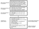
Figure. Diagnostic guidelines for clinicians and microbiologists in developed countries and resource-limited settings. 1) The antimicrobial drug–susceptibility pattern can be useful for distinguishing Burkholderia pseudomallei (usually resistant to aminoglycosides and colistin or...
In many regions of the world, the lack of microbiology laboratories hampers the diagnosis of bacterial infections in general, including melioidosis. Nonetheless, where microbiology facilities exist, identification of patients with melioidosis can still be problematic because of the lack of awareness among clinicians and laboratory staff, including lack of awareness of low-level endemicity where indigenous cases have been described (e.g., in India and Brazil) and failure to elicit or communicate a history of travel from patients returning from melioidosis-endemic areas. Table 2 describes common misconceptions and pitfalls that can occur when diagnosing melioidosis; the Figure illustrates when to suspect melioidosis, what specimens to take, and what types of tests are available.
In melioidosis-endemic areas, if melioidosis is suspected, empiric treatment with antimicrobial agents effective against B. pseudomallei should be initiated immediately, before diagnostic results are available, in an effort to reduce the number of deaths. Diagnostic tests used in melioidosis-endemic areas should be able to confirm melioidosis with high accuracy, with high positive and negative predictive values. High positive and negative predictive values are essential if the test result is being used to determine whether melioidosis-specific antimicrobial agents (rather than broad-spectrum empirically used antimicrobial agents to cover melioidosis-specific and other pathogenic organisms) are appropriate and whether the patients need to be treated with prolonged oral therapy to prevent melioidosis relapse. A rapid test that could be used at the point of care would be most useful in melioidosis-endemic areas. The ideal rapid test should use inexpensive commonly available equipment, supplies, and reagents. It should require minimal training, be robust in a variety of laboratory conditions (temperature, humidity), and have a long shelf life. It should be accurate and reliable even when performed on direct specimens, to minimize the hazard of working with pure culture.
In areas where melioidosis is less common or in non–melioidosis-endemic areas, empiric antimicrobial therapy for acute sepsis may not include drugs active against B. pseudomallei. In addition, the positive predictive values of rapid tests are probably much lower because of the low-prevalence setting. Therefore, diagnostic tests developed for these regions should focus on methods that detect pathogens more broadly and include B. pseudomallei, such as 16S sequencing or multiplexed real-time PCR assays. A combination of antigen and antibody detection to provide high specificity and sensitivity might be a possible solution for this setting. Educating technicians and clinicians about diagnosis of melioidosis is also necessary. Reporting of cases that occur in areas where melioidosis is less common or in non–melioidosis-endemic areas might help familiarize technicians and clinicians with this pathogen and alert public health officials to potential outbreaks.
The timely and accurate diagnosis of melioidosis is needed to ensure that effective antimicrobial therapy is initiated or continued appropriately. Distinct diagnostic obstacles exist in settings where melioidosis is or is not endemic and in environments with low or high levels of resources. Common misconceptions and pitfalls relating to diagnostic microbiology can also hinder early detection. Efforts to culture B. pseudomallei from persons suspected to have melioidosis are paramount and should include culturing of all available specimens by using selective media such as Ashdown agar or B. cepacia agar. The need to make latex agglutination testing available for rapid identification of isolates, particularly in low-resource melioidosis-endemic areas, received widespread support. Simple point-of-care tests such as the LFI may become available in the near future and would enable rapid identification of isolates and direct detection in clinical specimens. This capacity will greatly aid rapid diagnosis in developed countries and in low-resource settings.
Dr Hoffmaster is the team lead of the Zoonoses and Select Agent Laboratory in the Bacterial Special Pathogens Branch, Division of High-Consequence Pathogens and Pathology, National Center for Emerging and Zoonotic Infectious Diseases, Centers for Disease Control and Prevention. His research interests include diagnostics and molecular epidemiology of numerous pathogens, including B. pseudomallei.
Acknowledgments
We thank Bart Currie, Mindy Glass Elrod, Rebecca Lipsitz, Rosemarie Aurigemma, Alec Ritchie, and Maureen Beanan for helpful discussions on this workshop’s agenda and speaker selection; Dennis Dixon, Suman Mukhopadhyay, and Thames Pickett for contributing to discussions during the workshop; and Yolanda Gaines and Kimberly Tutt for assistance with organizing this workshop.
The workshop was funded by the US Centers for Disease Control and Prevention. Names of vendors or manufacturers are provided as examples of available product sources; inclusion does not imply endorsement of the vendors, manufacturers, or products by the US Centers for Disease Control and Prevention or the US Department of Health and Human Services.
D.A. and R.H. are principal investigators and receive funding from Phase 1 US Department of Health and Human Services Small Business Technology Transfer award no. R41AI102482-02. R.H. is an employee of InBios International but does not have a stake in the company.
References
- Wiersinga WJ, Currie BJ, Peacock SJ. Melioidosis. N Engl J Med. 2012;367:1035–44. DOIPubMedGoogle Scholar
- Ngauy V, Lemeshev Y, Sadkowski L, Crawford G. Cutaneous melioidosis in a man who was taken as a prisoner of war by the Japanese during World War II. J Clin Microbiol. 2005;43:970–2. DOIPubMedGoogle Scholar
- Peacock SJ, Schweizer HP, Dance DA, Smith TL, Gee JE, Wuthiekanun V, Management of accidental laboratory exposure to Burkholderia pseudomallei and B. mallei. Emerg Infect Dis. 2008;14:e2. DOIPubMedGoogle Scholar
- John TJ, Jesudason MV, Lalitha MK, Ganesh A, Mohandas V, Cherian T, Melioidosis in India: the tip of the iceberg? Indian J Med Res. 1996;103:62–5 .PubMedGoogle Scholar
- Kibbler CC, Roberts CM, Ridgway GL, Spiro SG. Melioidosis in a patient from Bangladesh. Postgrad Med J. 1991;67:764–6. DOIPubMedGoogle Scholar
- Stewart T, Engelthaler DM, Blaney DD, Tuanyok A, Wangsness E, Smith TL, Epidemiology and investigation of melioidosis, southern Arizona. Emerg Infect Dis. 2011;17:1286–8. DOIPubMedGoogle Scholar
- Zehnder AM, Hawkins MG, Koski MA, Lifland B, Byrne BA, Swanson AA, Burkholderia pseudomallei isolates in 2 pet iguanas, California, USA. Emerg Infect Dis. 2014;20:304–6. DOIPubMedGoogle Scholar
- Wuthiekanun V, Dance DA, Wattanagoon Y, Supputtamongkol Y, Chaowagul W, White NJ. The use of selective media for the isolation of Pseudomonas pseudomallei in clinical practice. J Med Microbiol. 1990;33:121–6. DOIPubMedGoogle Scholar
- Wuthiekanun V, Suputtamongkol Y, Simpson AJ, Kanaphun P, White NJ. Value of throat swab in diagnosis of melioidosis. J Clin Microbiol. 2001;39:3801–2. DOIPubMedGoogle Scholar
- Cheng AC, Wuthiekanun V, Limmathurosakul D, Wongsuvan G, Day NP, Peacock SJ. Role of selective and nonselective media for isolation of Burkholderia pseudomallei from throat swabs of patients with melioidosis. J Clin Microbiol. 2006;44:2316. DOIPubMedGoogle Scholar
- Peacock SJ, Chieng G, Cheng AC, Dance DA, Amornchai P, Wongsuvan G, Comparison of Ashdown’s medium, Burkholderia cepacia medium, and Burkholderia pseudomallei selective agar for clinical isolation of Burkholderia pseudomallei. J Clin Microbiol. 2005;43:5359–61 and. DOIPubMedGoogle Scholar
- Thorn P, Krause V, Boutlis C, Currie B. Melioidosis in the Top End. The Northern Territory Disease Control Bulletin. 2005;12:1–2 [cited 2014 Dec 4]. http://www.health.nt.gov.au/library/scripts/objectifyMedia.aspx?file=pdf/11/41.pdf&siteID=1&str_title=Bulletin+December+2005.pdf
- Limmathurotsakul D, Wuthiekanun V, Chierakul W, Cheng AC, Maharjan B, Chaowagul W, Role and significance of quantitative urine cultures in diagnosis of melioidosis. J Clin Microbiol. 2005;43:2274–6. DOIPubMedGoogle Scholar
- Centers for Disease Control and Prevention. Laboratory exposure to Burkholderia pseudomallei—Los Angeles, California, 2003. MMWR Morb Mortal Wkly Rep. 2004;53:988–90 .PubMedGoogle Scholar
- Pumpuang A, Chantratita N, Wikraiphat C, Saiprom N, Day NP, Peacock SJ, Survival of Burkholderia pseudomallei in distilled water for 16 years. Trans R Soc Trop Med Hyg. 2011;105:598–600. DOIPubMedGoogle Scholar
- Yabuuchi E, Wang L, Arakawa M, Yano I. Survival of Pseudomonas pseudomallei strains at 5 degrees C. Kansenshogaku Zasshi. 1993;67:331–5. DOIPubMedGoogle Scholar
- Larsen E, Smith JJ, Norton R, Corkeron M. Survival, sublethal injury, and recovery of environmental Burkholderia pseudomallei in soil subjected to desiccation. Appl Environ Microbiol. 2013;79:2424–7. DOIPubMedGoogle Scholar
- Holland DJ, Wesley A, Drinkovic D, Currie BJ. Cystic fibrosis and Burkholderia pseudomallei: an emerging problem? Clin Infect Dis. 2002;35:e138–40. DOIPubMedGoogle Scholar
- Wesley A, Holland D, Currie B. Burkholderia pseudomallei in two siblings with cystic fibrosis and evidence of likely person to person transmission in the family setting. In: Proceedings of Fourth Australian and New Zealand Cystic Fibrosis Conference; 2001 Aug 23–25, Brisbane, Australia. p. 46.
- Price EP, Sarovich DS, Mayo M, Tuanyok A, Drees KP, Kaestli M, Within-host evolution of Burkholderia pseudomallei over a twelve-year chronic carriage infection. MBiol. 2013;4:e00388–13.PubMedGoogle Scholar
- O’Sullivan BP, Torres B, Conidi G, Smole S, Gauthier C, Stauffer KE, Burkholderia pseudomallei infection in a child with cystic fibrosis: acquisition in the Western Hemisphere. Chest. 2011;140:239–42. DOIPubMedGoogle Scholar
- Huis in ’t Veld D, Wuthiekanun V, Cheng AC, Chierakul W, Chaowagul W, Brouwer AE, The role and significance of sputum cultures in the diagnosis of melioidosis. Am J Trop Med Hyg. 2005;73:657–61 .PubMedGoogle Scholar
- Limmathurotsakul D, Jamsen K, Arayawichanont A, Simpson JA, White LJ, Lee SJ, Defining the true sensitivity of culture for the diagnosis of melioidosis using Bayesian latent class models. PLoS ONE. 2010;5:e12485. DOIPubMedGoogle Scholar
- Roesnita B, Tay ST, Puthucheary SD, Sam IC. Diagnostic use of Burkholderia pseudomallei selective media in a low prevalence setting. Trans R Soc Trop Med Hyg. 2012;106:131–3. DOIPubMedGoogle Scholar
- Glass MB, Beesley CA, Wilkins PP, Hoffmaster AR. Comparison of four selective media for the isolation of Burkholderia mallei and Burkholderia pseudomallei. Am J Trop Med Hyg. 2009;80:1023–8 .PubMedGoogle Scholar
- Wongsuvan G, Limmathurotsakul D, Wannapasni S, Chierakul W, Teerawattanasook N, Wuthiekanun V. Lack of correlation of Burkholderia pseudomallei quantities in blood, urine, sputum and pus. Southeast Asian J Trop Med Public Health. 2009;40:781–4 .PubMedGoogle Scholar
- Dance DA, Wuthiekanun V, Naigowit P, White NJ. Identification of Pseudomonas pseudomallei in clinical practice: use of simple screening tests and API 20NE. J Clin Pathol. 1989;42:645–8. DOIPubMedGoogle Scholar
- Tandhavanant S, Wongsuvan G, Wuthiekanun V, Teerawattanasook N, Day NP, Limmathurotsakul D, Monoclonal antibody-based immunofluorescence microscopy for the rapid identification of Burkholderia pseudomallei in clinical specimens. Am J Trop Med Hyg. 2013;89:165–8. DOIPubMedGoogle Scholar
- Inglis TJ, Chiang D, Lee GS, Chor-Kiang L. Potential misidentification of Burkholderia pseudomallei by API 20NE. Pathology. 1998;30:62–4. DOIPubMedGoogle Scholar
- Lowe P, Engler C, Norton R. Comparison of automated and nonautomated systems for identification of Burkholderia pseudomallei. J Clin Microbiol. 2002;40:4625–7. DOIPubMedGoogle Scholar
- Inglis TJ, Merritt A, Chidlow G, Aravena-Roman M, Harnett G. Comparison of diagnostic laboratory methods for identification of Burkholderia pseudomallei. J Clin Microbiol. 2005;43:2201–6. DOIPubMedGoogle Scholar
- Glass MB, Popovic T. Preliminary evaluation of the API 20NE and RapID NF plus systems for rapid identification of Burkholderia pseudomallei and B. mallei. J Clin Microbiol. 2005;43:479–83. DOIPubMedGoogle Scholar
- Amornchai P, Chierakul W, Wuthiekanun V, Mahakhunkijcharoen Y, Phetsouvanh R, Currie BJ, Accuracy of Burkholderia pseudomallei identification using the API 20NE system and a latex agglutination test. J Clin Microbiol. 2007;45:3774–6. DOIPubMedGoogle Scholar
- Koh TH, Yong Ng LS, Foon Ho JL, Sng LH, Wang GC, Tzer Pin Lin RV. Automated identification systems and Burkholderia pseudomallei. J Clin Microbiol. 2003;41:1809. DOIPubMedGoogle Scholar
- Weissert C, Dollenmaier G, Rafeiner P, Riehm J, Schultze D. Burkholderia pseudomallei misidentified by automated system. Emerg Infect Dis. 2009;15:1799–801. DOIPubMedGoogle Scholar
- Lowe P, Haswell H, Lewis K. Use of various common isolation media to evaluate the new VITEK 2 colorimetric GN card for identification of Burkholderia pseudomallei. J Clin Microbiol. 2006;44:854–6. DOIPubMedGoogle Scholar
- Zong Z, Wang X, Deng Y, Zhou T. Misidentification of Burkholderia pseudomallei as Burkholderia cepacia by the VITEK 2 system. J Med Microbiol. 2012;61:1483–4. DOIPubMedGoogle Scholar
- Podin Y, Kaestli M, McMahon N, Hennessy J, Ngian HU, Wong JS, Reliability of automated biochemical identification of Burkholderia pseudomallei is regionally dependent. J Clin Microbiol. 2013;51:3076–8. DOIPubMedGoogle Scholar
- Hodgson K, Engler C, Govan B, Ketheesan N, Norton R. Comparison of routine bench and molecular diagnostic methods in identification of Burkholderia pseudomallei. J Clin Microbiol. 2009;47:1578–80. DOIPubMedGoogle Scholar
- Podin Y, Sarovich DS, Price EP, Kaestli M, Mayo M, Hii K, Burkholderia pseudomallei isolates from Sarawak, Malaysian Borneo, are predominantly susceptible to aminoglycosides and macrolides. Antimicrob Agents Chemother. 2014;58:162–6. DOIPubMedGoogle Scholar
- Anuntagool N, Naigowit P, Petkanchanapong V, Aramsri P, Panichakul T, Sirisinha S. Monoclonal antibody-based rapid identification of Burkholderia pseudomallei in blood culture fluid from patients with community-acquired septicaemia. J Med Microbiol. 2000;49:1075–8 .PubMedGoogle Scholar
- Dharakul T, Songsivilai S, Smithikarn S, Thepthai C, Leelaporn A. Rapid identification of Burkholderia pseudomallei in blood cultures by latex agglutination using lipopolysaccharide-specific monoclonal antibody. Am J Trop Med Hyg. 1999;61:658–62 .PubMedGoogle Scholar
- Ekpo P, Rungpanich U, Pongsunk S, Naigowit P, Petkanchanapong V. Use of protein-specific monoclonal antibody-based latex agglutination for rapid diagnosis of Burkholderia pseudomallei infection in patients with community-acquired septicemia. Clin Vaccine Immunol. 2007;14:811–2. DOIPubMedGoogle Scholar
- Kiratisin P, Santanirand P, Chantratita N, Kaewdaeng S. Accuracy of commercial systems for identification of Burkholderia pseudomallei versus Burkholderia cepacia. Diagn Microbiol Infect Dis. 2007;59:277–81 . DOIPubMedGoogle Scholar
- Lowe W, March JK, Bunnell AJ, O’Neill KL, Robison RA. PCR-based methodologies used to detect and differentiate the Burkholderia pseudomallei complex: B. pseudomallei, B. mallei, and B. thailandensis. Curr Issues Mol Biol. 2013;16:23–54 .PubMedGoogle Scholar
- Price EP, Dale JL, Cook JM, Sarovich DS, Seymour ML, Ginther JL, Development and validation of Burkholderia pseudomallei–specific real-time PCR assays for clinical, environmental or forensic detection applications. PLoS ONE. 2012;7:e37723. DOIPubMedGoogle Scholar
- Novak RT, Glass MB, Gee JE, Gal D, Mayo MJ, Currie BJ, Development and evaluation of a real-time PCR assay targeting the type III secretion system of Burkholderia pseudomallei. J Clin Microbiol. 2006;44:85–90. DOIPubMedGoogle Scholar
- Morse SA, Kellogg RB, Perry S, Meyer RF, Bray D, Nichelson D, Detecting biothreat agents: the Laboratory Response Network. ASM News. 2003;69:433–7.
- Gee JE, Sacchi CT, Glass MB, De BK, Weyant RS, Levett PN, Use of 16S rRNA gene sequencing for rapid identification and differentiation of Burkholderia pseudomallei and B. mallei. J Clin Microbiol. 2003;41:4647–54 . DOIPubMedGoogle Scholar
- Clinical and Laboratory Standards Institute. Methods for antimicrobial dilution and disk susceptibility testing of infrequently isolated or fastidious bacteria. CLSI document M45–A2. Wayne (PA): The Institute; 2010.
- Wuthiekanun V, Cheng AC, Chierakul W, Amornchai P, Limmathurotsakul D, Chaowagul W, Trimethoprim/sulfamethoxazole resistance in clinical isolates of Burkholderia pseudomallei. J Antimicrob Chemother. 2005;55:1029–31. DOIPubMedGoogle Scholar
- Jenney AW, Lum G, Fisher DA, Currie BJ. Antibiotic susceptibility of Burkholderia pseudomallei from tropical northern Australia and implications for therapy of melioidosis. Int J Antimicrob Agents. 2001;17:109–13. DOIPubMedGoogle Scholar
- Piliouras P, Ulett GC, Ashhurst-Smith C, Hirst RG, Norton RE. A comparison of antibiotic susceptibility testing methods for cotrimoxazole with Burkholderia pseudomallei. Int J Antimicrob Agents. 2002;19:427–9. DOIPubMedGoogle Scholar
- Chantratita N, Tandhavanant S, Wongsuvan G, Wuthiekanun V, Teerawattanasook N, Day NP, Rapid detection of Burkholderia pseudomallei in blood cultures using a monoclonal antibody–based immunofluorescent assay. Am J Trop Med Hyg. 2013;89:971–2. DOIPubMedGoogle Scholar
- Wuthiekanun V, Desakorn V, Wongsuvan G, Amornchai P, Cheng AC, Maharjan B, Rapid immunofluorescence microscopy for diagnosis of melioidosis. Clin Diagn Lab Immunol. 2005;12:555–6 .PubMedGoogle Scholar
- Thibault FM, Valade E, Vidal DR. Identification and discrimination of Burkholderia pseudomallei, B. mallei, and B. thailandensis by real-time PCR targeting type III secretion system genes. J Clin Microbiol. 2004;42:5871–4 . DOIPubMedGoogle Scholar
- Kaestli M, Richardson LJ, Colman RE, Tuanyok A, Price EP, Bowers JR, Comparison of TaqMan PCR assays for detection of the melioidosis agent Burkholderia pseudomallei in clinical specimens. J Clin Microbiol. 2012;50:2059–62. DOIPubMedGoogle Scholar
- Meumann EM, Novak RT, Gal D, Kaestli ME, Mayo M, Hanson JP, Clinical evaluation of a type III secretion system real-time PCR assay for diagnosing melioidosis. J Clin Microbiol. 2006;44:3028–30. DOIPubMedGoogle Scholar
- Chantratita N, Meumann E, Thanwisai A, Limmathurotsakul D, Wuthiekanun V, Wannapasni S, Loop-mediated isothermal amplification method targeting the TTS1 gene cluster for the detection of Burkholderia pseudomallei and diagnosis of melioidosis. J Clin Microbiol. 2008;46:568–73 . DOIPubMedGoogle Scholar
- Houghton RL, Reed DE, Hubbard MA, Dillon MJ, Chen H, Currie BJ, Development of a prototype lateral flow immunoassay (LFI) for the rapid diagnosis of melioidosis. PLoS Negl Trop Dis. 2014;8:e2727. DOIPubMedGoogle Scholar
- Sim BM, Chantratita N, Ooi WF, Nandi T, Tewhey R, Wuthiekanun V, Genomic acquisition of a capsular polysaccharide virulence cluster by non-pathogenic Burkholderia isolates. Genome Biol. 2010;11:R89. DOIPubMedGoogle Scholar
- Cheng AC, O’Brien M, Freeman K, Lum G, Currie BJ. Indirect hemagglutination assay in patients with melioidosis in northern Australia. Am J Trop Med Hyg. 2006;74:330–4 .PubMedGoogle Scholar
- Cheng AC, Peacock SJ, Limmathurotsakul D, Wongsuvan G, Chierakul W, Amornchai P, Prospective evaluation of a rapid immunochromogenic cassette test for the diagnosis of melioidosis in northeast Thailand. Trans R Soc Trop Med Hyg. 2006;100:64–7. DOIPubMedGoogle Scholar
- Harris PN, Ketheesan N, Owens L, Norton RE. Clinical features that affect indirect-hemagglutination-assay responses to Burkholderia pseudomallei. Clin Vaccine Immunol. 2009;16:924–30. DOIPubMedGoogle Scholar
- Armstrong PK, Anstey NM, Kelly PM, Currie BJ, Martins N, Dasari P, Seroprevalence of Burkholderia pseudomallei in East Timorese refugees: implications for healthcare in East Timor. Southeast Asian J Trop Med Public Health. 2005;36:1496–502 .PubMedGoogle Scholar
- Clayton AJ, Lisella RS, Martin DG. Melioidosis: a serological survey in military personnel. Mil Med. 1973;138:24–6 .PubMedGoogle Scholar
- Chantratita N, Wuthiekanun V, Thanwisai A, Limmathurotsakul D, Cheng AC, Chierakul W, Accuracy of enzyme-linked immunosorbent assay using crude and purified antigens for serodiagnosis of melioidosis. Clin Vaccine Immunol. 2007;14:110–3. DOIPubMedGoogle Scholar
- Limmathurotsakul D, Chantratita N, Teerawattanasook N, Piriyagitpaiboon K, Thanwisai A, Wuthiekanun V, Enzyme-linked immunosorbent assay for the diagnosis of melioidosis: better than we thought. Clin Infect Dis. 2011;52:1024–8. DOIPubMedGoogle Scholar
- Trikalinos TA, Balion CM. Chapter 9: options for summarizing medical test performance in the absence of a “gold standard.”. J Gen Intern Med. 2012;27(Suppl 1):S67–75. DOIPubMedGoogle Scholar
- Lim C, Wannapinij P, White L, Day NP, Cooper BS, Peacock SJ, Using a web-based application to define the accuracy of diagnostic tests when the gold standard is imperfect. PLoS ONE. 2013;8:e79489. DOIPubMedGoogle Scholar
- Karger A, Stock R, Ziller M, Elschner MC, Bettin B, Melzer F, Rapid identification of Burkholderia mallei and Burkholderia pseudomallei by intact cell matrix-assisted laser desorption/ionisation mass spectrometric typing. BMC Microbiol. 2012;12:229. DOIPubMedGoogle Scholar
- Weigel LM, Sue D, Michel PA, Kitchel B, Pillai SP. A rapid antimicrobial susceptibility test for Bacillus anthracis. Antimicrob Agents Chemother. 2010;54:2793–800 . DOIPubMedGoogle Scholar
- Chetchotisakd P, Chierakul W, Chaowagul W, Anunnatsiri S, Phimda K, Mootsikapun P, Trimethoprim-sulfamethoxazole versus trimethoprim-sulfamethoxazole plus doxycycline as oral eradicative treatment for melioidosis (MERTH): a multicentre, double-blind, non-inferiority, randomised controlled trial. Lancet. 2014;383:807–14 . DOIPubMedGoogle Scholar
Figure
Tables
Suggested citation for this article: Hoffmaster AR, AuCoin D, Baccam P, Baggett HC, Baird R, Bhengsri S, et al. Melioidosis Diagnostic Workshop, 2013. Emerg Infect Dis. 2015 Feb [date cited]. http://dx.doi.org/10.3201/eid2102.141045
1Held September 14, 2013, in Bangkok, Thailand.
Table of Contents – Volume 21, Number 2—February 2015
| EID Search Options |
|---|
|
|
|
|
|
|
Please use the form below to submit correspondence to the authors or contact them at the following address:
Alex R. Hoffmaster, Centers for Disease Control and Prevention, 1600 Clifton Rd NE, Mailstop G34, Atlanta, GA 30329-4027
Top