Volume 21, Number 5—May 2015
Dispatch
Antimicrobial Drug Resistance of Vibrio cholerae, Democratic Republic of the Congo
Cite This Article
Citation for Media
Abstract
We analyzed 1,093 Vibrio cholerae isolates from the Democratic Republic of the Congo during 1997–2012 and found increasing antimicrobial drug resistance over time. Our study also demonstrated that the 2011–2012 epidemic was caused by an El Tor variant clonal complex with a single antimicrobial drug susceptibility profile.
Cholera is an acute intestinal infection caused by Vibrio cholerae (1). Although hydration remains the primary treatment for cholera, antimicrobial drug therapy is recommended for severely ill patients (2). However, multidrug-resistant V. cholerae strains have long been observed in Africa (3), and strains exhibiting new resistance phenotypes have emerged during recent epidemics (4). It is therefore critical to carefully monitor changes in strains’ susceptibility to antimicrobial drugs in each African country and adapt treatment recommendations accordingly.
Few longitudinal studies assessing shifts in the resistance of V. cholerae to antimicrobial drugs in Africa have been established. The available studies are limited either to a restricted area (5) or a short time period (6). We describe the long-term evolution of antimicrobial drug susceptibility of an extensive set of V. cholerae isolates collected in the Democratic Republic of the Congo (DRC). We applied whole-genome sequencing and multiple locus variable-number tandem-repeat analysis (MLVA) to clarify the mechanisms behind the aggressive epidemic of 2011–2012 that spread throughout the country, affecting regions to which cholera was not endemic (7).
Sample collection included all available isolates from major outbreaks in the DRC during 1997–2012, which were stored at the National Institute of Biomedical Research. The Table shows the locations where the 1,093 tested isolates were collected.
V. cholerae O1 strains stored in nutrient agar during 1997–2012 were cultured on thiosulfate citrate bile salts agar and nutrient agar at the National Laboratory, Kinshasa, DRC. The strains were enriched in alkaline peptone water liquid medium and incubated at 37°C for 18–24 h. Biochemical and serogroup characterization was subsequently performed according to standard protocols (8).
Antimicrobial drug susceptibility testing of 1,093 confirmed V. cholerae isolates was performed by using the Kirby-Bauer disk diffusion method (9) with Mueller agar (bioMérieux, Marcy l’Etoile, France). V. cholerae O1 Ogawa and Inaba reference strains (ATCC) served as controls. Isolates were tested against 9 antimicrobial drugs as follows: ampicillin (10 µg), chloramphenicol (30 µg), sulfamethoxazole/trimethoprim (1.25 + 23.75 µg), tetracycline (30 µg), doxycycline (30 µg), norfloxacin (5 µg), ciprofloxacin (5 µg), nalidixic acid (30 µg), and erythromycin (30 µg) (bioMérieux). Interpretation of inhibition diameters (sensitive, intermediate, and resistant) was performed according to Clinical and Laboratory Standards Institute guidelines (10). When no interpretive criteria for V. cholerae were available from these guidelines, breakpoints for Enterobacteriaceae were applied by using Escherichia coli ATCC 25922 for quality control. The few intermediate results were categorized as resistant for this study. The strains were then regrouped into 21 resistance profiles.
Seventy-four clinical isolates from the 2011–2012 epidemic, spatiotemporally representative of outbreak diffusion, were subcultured and transported to Marseille, France. In Marseille, the strains were recultivated and identified as previously described (11). For DNA extraction, a 50-colony aliquot of cultured cells was suspended in 500 µL NucliSENS easyMAG lysis buffer (bioMérieux). DNA was extracted by using a NucliSENS easyMAG platform (bioMérieux) according to the manufacturer’s instructions. MLVA-based genotyping of the V. cholerae isolates and eBURST analysis (http://eBURST.mlst.net) were performed as previously described (11). To perform a phylogenetic assessment of the core V. cholerae genome on the basis of genome-wide single nucleotide polymorphisms, whole-genome sequencing was performed on an isolate (L286) collected at epidemic onset by using a HiSeq Illumina System (Illumina, San Diego, CA, USA) as previously described (12).
Figure 1
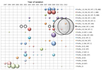
Figure 1. Vibrio cholerae strain antimicrobial drug resistance profiles plotted by year, Democratic Republic of the Congo, 1997–2012. On the basis of the antibiogram results, strains were grouped into 21 antimicrobial drug resistance...
Figure 2
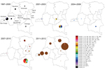
Figure 2. Spatiotemporal localization of isolate antimicrobial drug resistance profiles by time period and province, Democratic Republic of the Congo, 1997–2012. Strains were grouped into 21 antimicrobial drug resistance profiles. The antimicrobial drugs...
A spatiotemporal analysis was performed on the basis of the antibiogram profiles of V. cholerae isolates collected in the DRC during 1997–2012. The strain profiles were plotted by year (Figure 1) and then mapped by year and province. Using these data, we regrouped them into 5 representative periods (Figure 2). The V. cholerae strains displayed an increasingly complex resistance phenotype to various antimicrobial drugs. Sulfamethoxazole/trimethoprim resistance was observed initially, followed by resistance to nalidixic acid, erythromycin, and chloramphenicol during the early 2000s. Although sensitivity to fluoroquinolones seemed to be preserved, strain resistance patterns continued to evolve with the circulation of isolates resistant to tetracyclines and ampicillin from 2007–2010. Finally, isolates collected during 2011–2012, which was marked by the westward spread of a major epidemic (7), displayed a single antimicrobial drug susceptibility profile: resistance to most antimicrobial drugs except cyclines and fluoroquinolones.
Serotype analysis of the 1,093 V. cholerae isolates showed that Inaba strains were restricted to the western region of the country, Ogawa strains were isolated in the east and south, and Hikojima strains were restricted to Oriental Province, in the northeastern region of the country. During 2001–2010, Inaba and Ogawa serotypes were observed, but Ogawa predominated; during 2011–2012, these serotypes switched, and the Ogawa serotype was almost completely replaced by Inaba.
To examine the particular 2011–2012 epidemic that spread throughout the DRC (7), 74 V. cholerae isolates were assessed by using MLVA and eBURST analysis. Overall, the isolates displayed 19 different MLVA genotypes, of which 18 grouped into 1 clonal complex. eBURST analysis indicated that the clonal complex likely arose from a founder strain identified at the beginning of the epidemic. Furthermore, whole-genome sequence analysis of an isolate identified in March 2011 in Lubunga, Oriental Province (L286), revealed that the strain was an El Tor variant with CTX-3 type phage and a RS1 satellite phage. Phylogeny analysis situated this DRC strain close to the major Kenyan clade in the most recent wave of the seventh pandemic (data not shown).
Analysis of a panel of V. cholerae clinical isolates from the DRC from 1997–2012 highlighted a loss of sensitivity to leading antimicrobial drugs, although strains remain susceptible to fluoroquinolones. However, a risk for emergence and spread of fluoroquinolone-resistant strains exists, as has been shown elsewhere in Africa (13). Because resistance to nalidixic acid is frequently associated with decreased susceptibility to fluoroquinolones, nalidixic acid resistance must be detected to monitor the emergence of highly resistant strains (14).
Our findings also provide new insight regarding the cholera epidemic of 2011–2012. This epidemic appears to have been caused by the expansion of a specific V. cholerae subpopulation, which rapidly diffused countrywide. Furthermore, sequence analysis showed that the clone responsible for this epidemic, an El Tor variant with CTX-3 type phage, falls close to the major Kenyan clade in wave 3 of the seventh pandemic. This observation correlates with a 2011 study demonstrating that the seventh cholera pandemic had been caused by specific strains originating from a unique ancestral clone that have spread globally in successive waves (12). The 2011–2012 isolates displayed a specific antimicrobial drug resistance pattern, characterized by the return of tetracycline and doxycycline sensitivity. The outbreak strain also represented a serotype switch from Ogawa to Inaba. However, further MLVA genotyping of preoutbreak isolates is required to determine whether these strains were already present in the region or if they represent a new V. cholerae population.
This study demonstrates that molecular and microbiological analyses of V. cholerae isolates provide extensive insight into the mechanisms of cholera epidemics. MLVA and whole-genome sequencing are powerful tools for elucidating epidemic dynamics because these methods have been used to link distinct outbreaks and identify the origin of certain epidemic V. cholerae strains (15). Improved sampling of clinical isolates is essential to monitor changes in pathogen antimicrobial drug resistance and elucidate the dissemination pathways of toxigenic strains to ensure proper management of patients requiring antimicrobial drug treatment and to appropriately direct the public health response.
Ms. Miwanda is a biologist specializing in the diagnosis of bacterial diseases of epidemic potential and head of the Surveillance des Maladies à Potentiel Epidémique unit at the DRC Public Health National Laboratory. She is responsible for the diagnosis of meningitis, cholera, plague, and bloody diarrhea.
Acknowledgments
We thank Benoit Kebela Ilunga and Vital Mondonge Makuma for authorizing access to data and sharing their expertise. François-Xavier Mbopi- Kéou, Ditu Kazambu, Dieulla Delissaint, and Louis Koyange Delysogo assisted with the collection of V. cholerae isolates. We also thank the health authorities of the Provincial Directorates and the DRC health zone teams for authorizing and supporting the study.
The collection of strains and biological analyses were supported by many partners, including the World Health Organization, the Veolia Foundation, and the African Cholera Surveillance Network. Epidemiologic surveillance was financially supported by the World Health Organization, Epicentre, and the Belgian Development Cooperation.
References
- Centers for Disease Control and Prevention. Vibrio cholerae infection: antibiotic treatment. Recommendations for the use of antibiotics for the treatment of cholera. Atlanta: The Centers; 2013 [cited 2013 Aug 5]. http://www.cdc.gov/cholera/treatment/antibiotic-treatment.html
- Finch MJ, Morris JG Jr, Kaviti J, Kagwanja W, Levine MM. Epidemiology of antimicrobial resistant cholera in Kenya and East Africa. Am J Trop Med Hyg. 1988;39:484–90.PubMedGoogle Scholar
- Ngandjio A, Tejiokem M, Wouafo M, Ndome I, Yonga M, Guenole A, Antimicrobial resistance and molecular characterization of Vibrio cholerae O1 during the 2004 and 2005 outbreak of cholera in Cameroon. Foodborne Pathog Dis. 2009;6:49–56. DOIPubMedGoogle Scholar
- Mandomando I, Espasa M, Vallès X, Sacarlal J, Sigaúque B, Ruiz J, Antimicrobial resistance of Vibrio cholerae O1 serotype Ogawa isolated in Manhiça District Hospital, southern Mozambique. J Antimicrob Chemother. 2007;60:662–4. Epub 2007 Jul 11. DOIPubMedGoogle Scholar
- Materu SF, Lema OE, Mukunza HM, Adhiambo CG, Carter JY. Antibiotic resistance pattern of Vibrio cholerae and Shigella causing diarrhoea outbreaks in the eastern Africa region: 1994–1996. East Afr Med J. 1997;74:193–7.PubMedGoogle Scholar
- Bompangue D, Vesenbeckh SM, Giraudoux P, Castro M, Muyembe JJ, Kebela Ilunga B, et al. Cholera ante portas—the re-emergence of cholera in Kinshasa after a ten-year hiatus. PLoS Curr. 2012;4:RRN1310. PMID: 22453903
- Centers for Disease Control and Prevention. Laboratory methods for the diagnosis of epidemic dysentery and cholera. Atlanta: The Centers; 1999 [cited 2013 Apr 28]. http://www.cdc.gov/cholera/pdf/Laboratory-Methods-for-the-Diagnosis-of-Epidemic-Dysentery-and-Cholera.pdf
- Jorgensen JH, Turnidge JD. Susceptibility test methods: dilution and disk diffusion methods. In: Murray PR, Baron EJ, Jorgensen JH, Landry ML, Pfaller MA, editors. Manual of clinical microbiology. 9th ed. Washington (DC): ASM Press; 2007. p. 1152–72.
- Clinical and Laboratory Standards Institute. Performance standards for antimicrobial disk susceptibility tests; approved standard. 9th edition. Clinical and Laboratory Standards Institute document M2–A9. Wayne (PA): The Institute; 2006.
- Rebaudet S, Mengel MA, Koivogui L, Moore S, Mutreja A, Kande Y, Deciphering the origin of the 2012 cholera epidemic in Guinea by integrating epidemiological and molecular analyses. PLoS Negl Trop Dis. 2014;8:e2898. DOIPubMedGoogle Scholar
- Mutreja A, Kim DW, Thomson NR, Connor TR, Lee JH, Kariuki S, Evidence for several waves of global transmission in the seventh cholera pandemic. Nature. 2011;477:462–5. DOIPubMedGoogle Scholar
- Islam MS, Midzi SM , Charimari L, Cravioto A, Endtz HP. Susceptibility to fluoroquinolones of Vibrio cholerae O1 isolated from diarrheal patients in Zimbabwe. JAMA. 2009;302:2321–2. DOIPubMedGoogle Scholar
- Ray P, Sharma J, Marak R, Garg RK. Predictive efficacy of nalidixic acid resistance as a marker of fluoroquinolone resistance in Salmonella enterica var Typhi. Indian J Med Res. 2006;124:105–8.PubMedGoogle Scholar
- Hendriksen RS, Price LB, Schupp JM, Gillece JD, Kaas RS, Engelthaler DM, Population genetics of Vibrio cholerae from Nepal in 2010: evidence on the origin of the Haitian outbreak. MBio. 2011;2:e00157–11. DOIPubMedGoogle Scholar
Figures
Table
Cite This ArticleCrossRef reports the first page should be "e00157-11" not "e00157" in reference 15 "Hendriksen, Price, Schupp, Gillece, Kaas, Engelthaler, et al., 2011".
Table of Contents – Volume 21, Number 5—May 2015
| EID Search Options |
|---|
|
|
|
|
|
|
Please use the form below to submit correspondence to the authors or contact them at the following address:
Renaud Piarroux, APHM–Hôpital de la Timone, Laboratoire de Parasitologie-Mycologie, 264 rue Saint Pierre, 13385 Marseille CEDEX 05, France
Top