Detection of Circovirus in Foxes with Meningoencephalitis, United Kingdom, 2009–2013
Steve Bexton, Lidewij C. Wiersma, Sarah Getu, Peter R. van Run, Georges M.G.M. Verjans, Debby Schipper, Claudia M.E. Schapendonk, Rogier Bodewes, Lucy Oldroyd, Bart L. Haagmans, Marion M.P. Koopmans, and Saskia L. Smits

Author affiliations: RSPCA Norfolk Wildlife Hospital, East Winch, United Kingdom (S. Bexton); Erasmus Medical Center, Rotterdam, the Netherlands (L.C. Wiersma, S. Getu, P.R. van Run, G.M.G.M. Verjans, D. Schipper, C.M.E. Schapendonk, R. Bodewes, B.L. Haagmans, M.M.P. Koopmans, S.L. Smits); Abbey Veterinary Services, Newton Abbot, United Kingdom (L. Oldroyd); National Institute for Public Health and the Environment, Bilthoven, the Netherlands (M.M.P. Koopmans)
Main Article
Figure 2
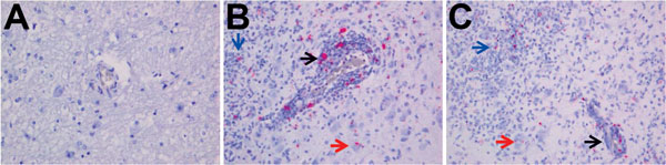
Figure 2. Detection of fox circovirus–specific transcripts in brain tissue of foxes with neurologic disease showing in situ hybridization of cerebrum with fox circovirus replication initiator protein gene–specific probe (original magnification ×200). A) Negative control fox VS7100012. The serum sample from this fox was negative for circovirus, and the animal did not exhibit signs of neurologic disease. B, C) Affected foxes VS7100005 and VS7100003, respectively. Both animals had neurologic disease, and their serum samples were positive for fox circovirus (see Table 1 for more information regarding these foxes). Black arrows indicate mononuclear cells in perivascular cuffs, blue arrows show inflammatory infiltrates in the neuropil, and red arrows point to staining in neuronal somata in cerebral gray matter of circovirus–positive animals with neurologic disease.
Main Article
Page created: June 15, 2015
Page updated: June 15, 2015
Page reviewed: June 15, 2015
The conclusions, findings, and opinions expressed by authors contributing to this journal do not necessarily reflect the official position of the U.S. Department of Health and Human Services, the Public Health Service, the Centers for Disease Control and Prevention, or the authors' affiliated institutions. Use of trade names is for identification only and does not imply endorsement by any of the groups named above.