Experimental Infection of Common Eider Ducklings with Wellfleet Bay Virus, a Newly Characterized Orthomyxovirus
Valerie Shearn-Bochsler

, Hon Sang Ip, Anne Ballmann, Jeffrey S. Hall, Andrew B. Allison, Jennifer Ballard, Julie C. Ellis, Robert Cook, Samantha E.J. Gibbs, and Chris Dwyer
Author affiliations: US Geological Survey National Wildlife Health Center, Madison, Wisconsin, USA (V. Shearn-Bochsler, H.S. Ip, A. Ballmann, J.S. Hall); Cornell University, Ithaca, New York, USA (A.B. Allison); Arkansas Game and Fish Commission, Little Rock, Arkansas, USA (J. Ballard); Tufts University, North Grafton, Massachusetts, USA (J.C. Ellis); National Park Service, Wellfleet, Massachusetts, USA (R. Cook); US Fish and Wildlife Service, Fort Collins, Colorado, USA (S.E.J. Gibbs); US Fish and Wildlife Service, Hadley, Massachusetts, USA (C. Dwyer)
Main Article
Figure 1
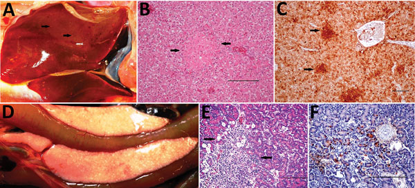
Figure 1. Gross, histopathologic, and immunohistochemical (IHC) findings in common eider (Somateria mollissima) ducklings experimentally infected with Wellfleet Bay virus (2 days postinoculation). A) Liver, enlarged, showing multifocal pinpoint areas of necrosis (arrows). B) Hematoxylin and eosin stain of liver tissue, showing focal hepatocellular necrosis (arrows). C) IHC stain of liver tissue, showing positive immunolabeling for Wellfleet Bay virus with multifocal staining of hepatocytes (arrows). D) Pancreas showing multifocal to coalescing acute necrosis. E) Hematoxylin and eosin stain of pancreas tissue, showing focally extensive necrosis of exocrine cells (arrows). F) IHC stain of pancreas tissue, showing positive immunolabeling for Wellfleet Bay virus in exocrine cells. Scale bars in panels B, C, E, and F indicate 100 μm.
Main Article
Page created: November 16, 2017
Page updated: November 16, 2017
Page reviewed: November 16, 2017
The conclusions, findings, and opinions expressed by authors contributing to this journal do not necessarily reflect the official position of the U.S. Department of Health and Human Services, the Public Health Service, the Centers for Disease Control and Prevention, or the authors' affiliated institutions. Use of trade names is for identification only and does not imply endorsement by any of the groups named above.