Skip directly to site content Skip directly to page options Skip directly to A-Z link Skip directly to A-Z link Skip directly to A-Z link
Volume 23, Number 12—December 2017
Research

Experimental Infection of Common Eider Ducklings with Wellfleet Bay Virus, a Newly Characterized Orthomyxovirus

Valerie Shearn-BochslerComments to Author , Hon Sang Ip, Anne Ballmann, Jeffrey S. Hall, Andrew B. Allison, Jennifer Ballard, Julie C. Ellis, Robert Cook, Samantha E.J. Gibbs, and Chris Dwyer
Author affiliations: US Geological Survey National Wildlife Health Center, Madison, Wisconsin, USA (V. Shearn-Bochsler, H.S. Ip, A. Ballmann, J.S. Hall); Cornell University, Ithaca, New York, USA (A.B. Allison); Arkansas Game and Fish Commission, Little Rock, Arkansas, USA (J. Ballard); Tufts University, North Grafton, Massachusetts, USA (J.C. Ellis); National Park Service, Wellfleet, Massachusetts, USA (R. Cook); US Fish and Wildlife Service, Fort Collins, Colorado, USA (S.E.J. Gibbs); US Fish and Wildlife Service, Hadley, Massachusetts, USA (C. Dwyer)

Main Article

Figure 2

Histopathologic and immunohistochemical (IHC) findings in lymphoid organs of control and infected common eider (Somateria mollissima) ducklings experimentally infected with Wellfleet Bay virus (WFBV) (2 days postinoculation). A) Hematoxylin and eosin (H&E) stain of thymic cortex tissue from a control duckling. B) H&E stain of thymic cortex tissue from an infected duckling, showing marked multifocal acute apoptosis of lymphocytes (arrows). C) IHC stain of thymus tissue from an infected du

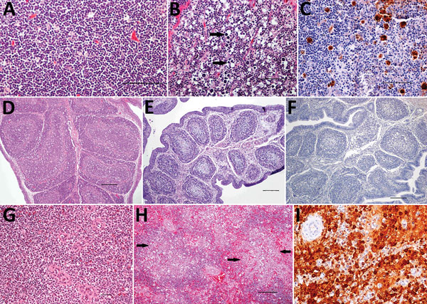
Figure 2. Histopathologic and immunohistochemical (IHC) findings in lymphoid organs of control and infected common eider (Somateria mollissima) ducklings experimentally infected with Wellfleet Bay virus (WFBV) (2 days postinoculation). A) Hematoxylin and eosin (H&E) stain of thymic cortex tissue from a control duckling. B) H&E stain of thymic cortex tissue from an infected duckling, showing marked multifocal acute apoptosis of lymphocytes (arrows). C) IHC stain of thymus tissue from an infected duckling, showing positive immunolabeling for WFBV in thymocytes. D) H&E stain of bursa of Fabricius tissue from a control duckling. E) H&E stain of bursa of Fabricius tissue from an infected duckling, showing marked diffuse lymphoid depletion. F) IHC stain of bursa of Fabricius tissue from an infected duckling showing negative immunolabeling for WFBV. G) H&E stain of spleen tissue from a control duckling. H) H&E stain of spleen tissue from an infected duckling, showing multifocal to coalescing acute necrosis (arrows). I) IHC stain of spleen tissue, showing positive immunolabeling for WFBV. I, original magnification ×200. Scale bars in panels A, B, and C indicate 50 µm and in panels D, E, F, G, and H indicate 100 µm.

Main Article

Page created: November 16, 2017
Page updated: November 16, 2017
Page reviewed: November 16, 2017
The conclusions, findings, and opinions expressed by authors contributing to this journal do not necessarily reflect the official position of the U.S. Department of Health and Human Services, the Public Health Service, the Centers for Disease Control and Prevention, or the authors' affiliated institutions. Use of trade names is for identification only and does not imply endorsement by any of the groups named above.
file_external