Volume 23, Number 12—December 2017
Synopsis
Fatal Outbreak in Tonkean Macaques Caused by Possibly Novel Orthopoxvirus, Italy, January 20151
Figure 4
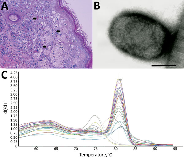
Figure 4. Results from necropsy of Tonkean macaque (Macaca tonkeana) from animal sanctuary, Italy, January 2015. A) Hematoxylin and eosin stain of cutaneous lesion. Focal epidermal necrosis, acanthosis ballooning degeneration, and acantholysis of keratinocytes was observed. Staining shows early vesiculation with eosinophilic intracytoplasmic inclusion bodies (arrows) in enlarged degenerated cells. B) Electron micrograph of skin lesion sample showing negatively stained brick-shaped viral particle of ≈160–220 nm, consistent with orthopoxvirus. Scale bar = 100 nm. C) SYBR Green (ThermoFisher Scientific, Waltham, MA, USA) real-time PCR melting curve of all tested samples. The y-axis shows the ratio of the change in fluorescence over the change in temperature. The average melting temperature (80.8°C ± 1°C) was consistent with that for the orthopoxvirus genome.
1Preliminary results from this study were presented at the Xth International Congress of the European Society for Veterinary Virology; August 31–September 3, 2015; Montpellier, France.
2These first authors were co–principal investigators who contributed equally to this article.