Postmortem Findings for 7 Neonates with Congenital Zika Virus Infection
Anastácio Q. Sousa

, Diane I.M. Cavalcante, Luciano M. Franco, Fernanda M.C. Araújo, Emília T. Sousa, José Telmo Valença-Junior, Dionne B. Rolim, Maria E.L. Melo, Pedro D.T. Sindeaux, Marialva T.F. Araújo, Richard D. Pearson, Mary E. Wilson, and Margarida M.L. Pompeu
Author affiliations: Federal University of Ceará, Fortaleza, Brazil (A.Q. Sousa, D.I.M. Cavalcante, L.M. Franco, E.T. Sousa, J.T. Valença-Junior, P.D.T. Sindeaux, M.M.L. Pompeu); Serviço de Verificação de Óbitos-SVO, Fortaleza (L.M. Franco, E.T. Sousa, J.T. Valença-Junior); Ceará State Central Public Health Laboratory, Fortaleza (F.M.C. Araújo, M.E.L. Melo); University of Fortaleza, Fortaleza (D.B. Rolim); Ceará State Secretariat of Health, Fortaleza (D.B. Rolim); Evandro Chagas Institute, Belém, Brazil (M.T.F. Araújo); University of Virginia School of Medicine, Charlottesville, Virginia, USA (R.D. Pearson); University of California, San Francisco, California, USA (M.E. Wilson); Harvard T.H. Chan School of Public Health, Boston, Massachusetts, USA (M.E. Wilson)
Main Article
Figure 1
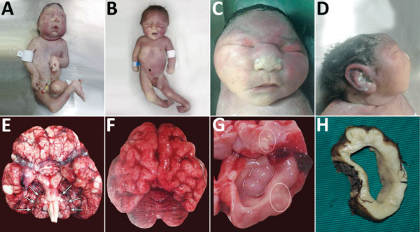
Figure 1. Physical signs in 4 of 7 neonates who died of congenital Zika virus infection, Brazil. A) Neonate 1: typical microcephaly phenotype; arthrogryposis in upper and lower limbs. B) Neonate 7: microcephaly without the typical microcephaly phenotype; arthrogryposis is also present. C) Neonate 3: typical microcephaly phenotype, with head circumference within reference limits, frontal view. D) Neonate 3: typical microcephaly phenotype, with head circumference within reference limits, profile view. E) Brain of neonate 2: symmetric cerebellar hypoplasia (arrows) and vascular congestion. F) Brain of neonate 3: pachygyria and severe vascular congestion. G) Brain of neonate 3: ventriculomegaly and macroscopic calcifications (circles). H) Brain of neonate 7: cross-section showing ventriculomegaly.
Main Article
Page created: June 19, 2017
Page updated: June 19, 2017
Page reviewed: June 19, 2017
The conclusions, findings, and opinions expressed by authors contributing to this journal do not necessarily reflect the official position of the U.S. Department of Health and Human Services, the Public Health Service, the Centers for Disease Control and Prevention, or the authors' affiliated institutions. Use of trade names is for identification only and does not imply endorsement by any of the groups named above.