Volume 23, Number 7—July 2017
Dispatch
Postmortem Findings for 7 Neonates with Congenital Zika Virus Infection
Figure 2
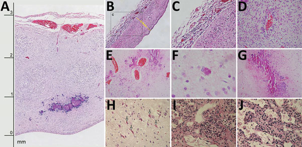
Figure 2. Histologic slides of tissues from 4 of 7 neonates who died of congenital Zika virus infection, Brazil. A) Neonate 1: severe cortical thinning (3 mm) with subventricular dystrophic calcification, reactive gliosis, and marked leptomeningeal congestion as well as marked depletion of neuronal precursors (original magnification ×10). B) Neonate 1: severe thinning of brain parenchyma (0.8 mm) with striking depletion of neuronal precursors (original magnification ×10). C) Neonate 1: lymphocytic leptomeningitis (enlargement of box in panel B; original magnification ×20). D) Neonate 6: white matter with lymphocytic perivascular cuffing and severe gliosis (original magnification ×40). E) Neonate 3: marked parenchymal vascular congestion and scattered coarse dystrophic calcification (original magnification ×20). F) Neonate 3: finely granular intracellular calcification (original magnification ×40). G) Neonate 7: band-like pattern of coarse dystrophic calcification at the junction of gray and white matter (original magnification ×10). H) Neonate 6: red neurons (arrows) in brain parenchyma (original magnification ×40). I) Neonate 1: focal interstitial lymphocytic pulmonary infiltrate (original magnification ×40). J) Neonate 1: expansion of alveolar septa with scattered lymphocytic and macrophage infiltrate (original magnification ×40).