Volume 24, Number 2—February 2018
Research
New Parvovirus Associated with Serum Hepatitis in Horses after Inoculation of Common Biological Product
Figure 4
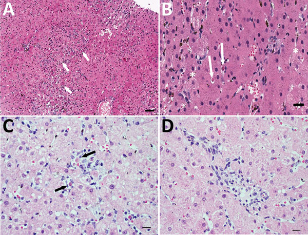
Figure 4. Histopathologic findings in the livers of 2 adult horses experimentally infected with an equine biological product containing equine parvovirus-hepatitis (EqPV-H). A) Liver biopsy sample from horse 1 obtained 82 days after inoculation with EqPV-H. Numerous individual and small clusters of lymphocytes are scattered about the parenchyma (arrows), indicative of lymphocytic lobular hepatitis. Hematoxylin and eosin (H&E) stain. Scale bar = 200 μm. B) Higher magnification image of the liver biopsy sample illustrated in panel A. Two individual necrotic hepatocytes are highlighted (arrows). Note the shrunken cell bodies, hypereosinophilic cytoplasm, and pyknotic nucleus, compatible with acidophil bodies. Other cells (asterisks) are swollen, with pale, mildly vacuolated cytoplasm, interpreted as hydropic degeneration. The cellular pleomorphism resulted in a mild degree of lobular disarray. Kupffer cells on the left side of the image contain hemosiderin, which is normal in horses. H&E stain. Scale bar = 50 μm. C) Liver biopsy sample from horse 2 obtained 100 days after inoculation with EqPV-H. Lymphocytes (black arrows) surround individual and small clusters of necrotic hepatocytes. Lymphocytic satellitosis implicates immune-mediated killing of hepatocytes by cytotoxic lymphocytes. H&E stain. Scale bar = 50 μm. D) Liver biopsy sample from horse 2. The portal tracts are infiltrated by small numbers of lymphocytes that breach the limiting plate and obscure the boundaries. No piecemeal necrosis (i.e., individual hepatocyte necrosis in the limiting plate) is detected. H&E stain. Scale bar = 50 μm.
1Deceased.