Volume 24, Number 2—February 2018
Research
Yersinia pestis Survival and Replication in Potential Ameba Reservoir
David W. Markman , Michael F. Antolin, Richard A. Bowen, William H. Wheat, Michael Woods, Mercedes Gonzalez-Juarrero, and Mary Jackson
Figure 4
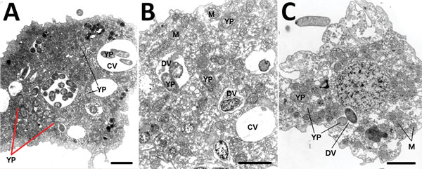
Figure 4. Representative transmission electron micrographs (TEM) depict Acanthamoeba castellanii amebae during A) 10-minute, B) 30-minute, and C) 24-hour co-cultures (multiplicity of infection 100) with Yersinia pestis (CO92 pgm+, pCD1, pGFPuv, amp+). Red arrows in panel A indicate potential intraameba mitotic division of Y. pestis bacterium. Visual analysis of TEM micrographs proved inconclusive for identifying the bacterial division septum. Y. pestis resides within the potential replicative niche of a tight-fitting vacuolar membrane, similar to Yersinia-containing vacuoles observed in macrophages. YP, Y. pestis; CV, central vacuole; DV, digestive vacuole; M, mitochondria. Scale bars indicate 3 μm.
Page created: January 17, 2018
Page updated: January 17, 2018
Page reviewed: January 17, 2018
The conclusions, findings, and opinions expressed by authors contributing to this journal do not necessarily reflect the official position of the U.S. Department of Health and Human Services, the Public Health Service, the Centers for Disease Control and Prevention, or the authors' affiliated institutions. Use of trade names is for identification only and does not imply endorsement by any of the groups named above.