Occupation-Associated Fatal Limbic Encephalitis Caused by Variegated Squirrel Bornavirus 1, Germany, 2013
Dennis Tappe

, Kore Schlottau, Daniel Cadar, Bernd Hoffmann, Lorenz Balke, Burkhard Bewig, Donata Hoffmann, Philip Eisermann, Helmut Fickenscher, Andi Krumbholz, Helmut Laufs, Monika Huhndorf, Maria Rosenthal, Walter Schulz-Schaeffer, Gabriele Ismer, Sven-Kevin Hotop, Mark Brönstrup, Anthonina Ott, Jonas Schmidt-Chanasit
1, and Martin Beer
1
Author affiliations: Bernhard Nocht Institute for Tropical Medicine, Hamburg, Germany (D. Tappe, D. Cadar, P. Eisermann, M. Rosenthal, J. Schmidt-Chanasit); Friedrich-Loeffler-Institut, Greifswald-Insel Riems, Germany (K. Schlottau, B. Hoffmann, D. Hoffmann, M. Beer); University Medical Center Schleswig-Holstein, Kiel, Germany (L. Balke, B. Bewig, H. Laufs, M. Huhndorf); Christian-Albrecht University of Kiel and University Medical Center, Kiel (H. Fickenscher, A. Krumbholz); Saarland University Medical Center, Homburg, Germany (W. Schulz-Schaeffer); Zoological Garden, Schleswig-Holstein, Germany (G. Ismer); Helmholtz Centre for Infection Research and German Centre for Infection Research, Braunschweig, Germany (S.-K. Hotop, M. Brönstrup); Euroimmun AG, Lübeck, Germany (A. Ott); German Centre for Infection Research, Hamburg (J. Schmidt-Chanasit)
Main Article
Figure 1

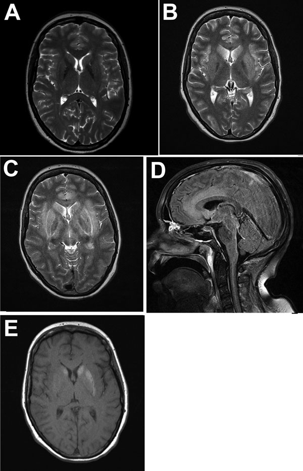
Figure 1. Magnetic resonance imaging of the brain throughout the course of the disease in patient who died of limbic encephalitis caused by variegated squirrel bornavirus 1 (VSBV-1), Germany, 2013. A) T2-weighted transversal image at admission showing no pathologic changes. B) T2-weighted image 3 weeks after admission showing edema in limbic structures (insula, hippocampus, anterior cingulate) and in the basal ganglia. C) T2-weighted image 4 weeks after admission showing progressive edema. Additional myelopathy extended from the medulla down into the thoracic segments (not shown). D) FLAIR image 4 weeks after admission showing edema in the anterior cingulate cortex. E) T1-weighted image 4 weeks after admission without contrast showing slight hemorrhage in the basal ganglia.
Main Article
Page created: May 18, 2018
Page updated: May 18, 2018
Page reviewed: May 18, 2018
The conclusions, findings, and opinions expressed by authors contributing to this journal do not necessarily reflect the official position of the U.S. Department of Health and Human Services, the Public Health Service, the Centers for Disease Control and Prevention, or the authors' affiliated institutions. Use of trade names is for identification only and does not imply endorsement by any of the groups named above.
