Volume 24, Number 7—July 2018
Synopsis
Typhus Group Rickettsiosis, Germany, 2010–20171
Figure 3
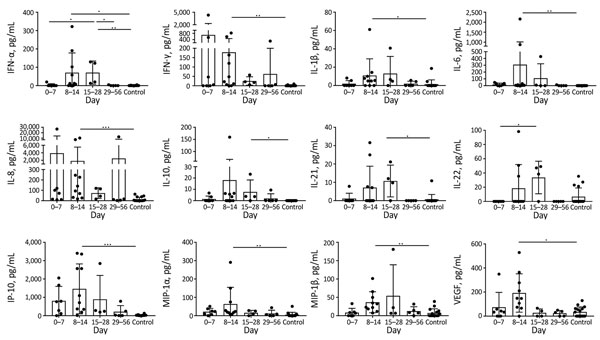
Figure 3. Cytokine and chemokine levels in serum from patients with imported and autochthonous typhus group rickettsiosis and controls, Germany, 2010–2017. Using a bead-based LEGENDplex assay (BioLegend, Fell, Germany), we analyzed 16 serum samples from healthy persons without rickettsial disease and 26 samples from 21 patients with typhus group rickettsiosis in parallel. We assigned 17 serum samples to the acute phase of illness (7 on days 0–7 and 10 on days 8–14), 4 to the prolonged phase, and 5 to the convalescent phase. Most serum cytokine levels started to increase in the first week of illness, peaked in the second week, and then started to decline again, except for IL-21 and IL-22, which reached their highest levels in the third week after symptom onset. Data are expressed as mean ± SD. Statistical analyses were performed by using the Kruskal-Wallis test and subsequently the Dunn multiple comparisons test. IFN, interferon; IL, interleukin; IP, interferon γ–induced protein; MIP, macrophage inflammatory protein; VEGF, vascular endothelial growth factor. Asterisks indicate statistically significant differences: *p<0.05; **p<0.01; ***p<0.001.
1Part of this study was orally presented at the annual meeting of the German Society for Hygiene and Microbiology, February 19–21, 2018, Bochum, Germany.