Volume 25, Number 1—January 2019
Synopsis
Clinical and Radiologic Characteristics of Human Metapneumovirus Infections in Adults, South Korea
Hyun Jung Koo, Han Na Lee, Sang-Ho Choi, Heungsup Sung, Hwa Jung Kim, and Kyung-Hyun Do
Figure 4
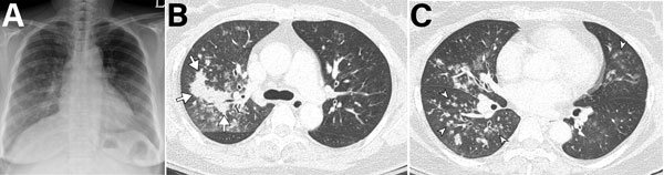
Figure 4. Imaging of 55-year-old immunocompetent woman with human metapneumovirus pneumonia, South Korea. A) Initial chest radiograph showing ill-defined patchy and nodular ground-glass opacities in the right lung and left lower lung zone. B, C) Chest computed tomography showing irregular nodular consolidation (arrows in panel B) and multiple ill-defined centrilobular nodular opacities (arrowheads in panel C) with mild bronchial wall thickening. Five days later, the lesions had resolved completely.
Page created: December 18, 2018
Page updated: December 18, 2018
Page reviewed: December 18, 2018
The conclusions, findings, and opinions expressed by authors contributing to this journal do not necessarily reflect the official position of the U.S. Department of Health and Human Services, the Public Health Service, the Centers for Disease Control and Prevention, or the authors' affiliated institutions. Use of trade names is for identification only and does not imply endorsement by any of the groups named above.