Skip directly to site content Skip directly to page options Skip directly to A-Z link Skip directly to A-Z link Skip directly to A-Z link
Volume 25, Number 4—April 2019
Research

Human-Origin Influenza A(H3N2) Reassortant Viruses in Swine, Southeast Mexico

Martha I. NelsonComments to Author , Carine K. Souza, Nídia S. Trovão, Andres Diaz, Ignacio Mena, Albert Rovira, Amy L. Vincent, Montserrat Torremorell, Douglas Marthaler1, and Marie R. Culhane
Author affiliations: National Institutes of Health, Bethesda, Maryland, USA (M.I. Nelson, N.S. Trovão); National Animal Disease Center, Ames, Iowa, USA (C.K. Souza, A.L. Vincent); Icahn School of Medicine at Mount Sinai, New York, New York, USA (N.S. Trovão, I. Mena); University of Minnesota, Saint Paul, Minnesota, USA (A. Diaz, A. Rovira, M. Torremorell, D. Marthaler, M.R. Culhane)

Main Article

Figure 4

Evolutionary relationships between H3 segments of avian influenza viruses collected in humans and swine globally. Time-scaled Bayesian maximum clade credibility tree is inferred for the H3 segment. The tree includes newly generated sequences from northwest and southeast Mexico, along with background sequences from swine in Mexico, Asia, the United States, and Canada, as well as seasonal H3N2 viruses from humans. The color of each branch indicates the most probable location state. Posterior proba

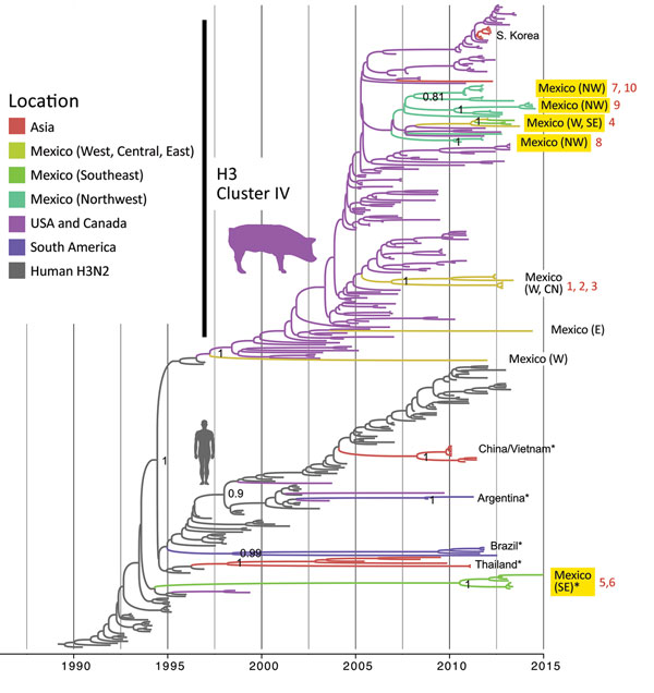
Figure 4. Evolutionary relationships between H3 segments of avian influenza viruses collected in humans and swine globally. Time-scaled Bayesian maximum clade credibility tree is inferred for the H3 segment. The tree includes newly generated sequences from northwest and southeast Mexico, along with background sequences from swine in Mexico, Asia, the United States, and Canada, as well as seasonal H3N2 viruses from humans. The color of each branch indicates the most probable location state. Posterior probabilities for key nodes are provided. Clades with viruses from swine in Mexico obtained for this study are highlighted in yellow. *Major virus introductions into a location, indicating direct introductions from humans. CN, central; NW, northwest; S, south; SE, southeast; W, west. The 10 H3N2 viruses selected for antigenic characterization (Figure 6) are indicated by numbers 1–10 in red. A more detailed phylogeny, including tip labels and all posterior probabilities, is provided in the Appendix Figure.

Main Article

1Current affiliation: Kansas State University, Manhattan, Kansas, USA.

Page created: March 17, 2019
Page updated: March 17, 2019
Page reviewed: March 17, 2019
The conclusions, findings, and opinions expressed by authors contributing to this journal do not necessarily reflect the official position of the U.S. Department of Health and Human Services, the Public Health Service, the Centers for Disease Control and Prevention, or the authors' affiliated institutions. Use of trade names is for identification only and does not imply endorsement by any of the groups named above.
file_external