Volume 26, Number 12—December 2020
CME ACTIVITY - Research
Clinical and Multimodal Imaging Findings and Risk Factors for Ocular Involvement in a Presumed Waterborne Toxoplasmosis Outbreak, Brazil1
Figure 3
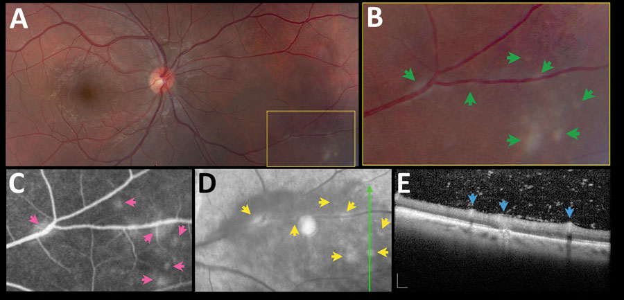
Figure 3. Asymptomatic retinochoroiditis in the right eye, detected in baseline examination (VA 0.0) of a 27-year-old man in a presumed waterborne toxoplasmosis outbreak, Brazil. A) Fundus photograph showing minimal vitreous haze; box indicates enlarged area on B); green arrows indicate multiple subtle and confluent gray-whitish punctate retinal infiltrates. C) Fundus fluorescein angiography. Pink arrows indicate leakage at the site of some of the punctate lesions. D) Fundus fluorescein angiography with red-free reflectance. Green line indicates site of optical coherence tomography scan. Yellow arrows indicate changes in the area of active focuses. E) Spectral-domain optical coherence tomography showing retinal thickening, and numerous overlying hyper-reflective dots. Blue arrows indicate multifocal hyper-reflectivity at the inner retinal layers. Scale bars indicate 200 µm. VA, visual acuity.
1Presented in part at the 2015 American Uveitis Society Fall meeting, November 15, 2014, Las Vegas, Nevada, USA