Volume 26, Number 12—December 2020
CME ACTIVITY - Research
Clinical and Multimodal Imaging Findings and Risk Factors for Ocular Involvement in a Presumed Waterborne Toxoplasmosis Outbreak, Brazil1
Camilo Brandão-de-Resende, Helena Hollanda Santos, Angel Alessio Rojas Lagos, Camila Munayert Lara, Jacqueline Souza Dutra Arruda, Ana Paula Maia Peixoto Marino, Lis Ribeiro do Valle Antonelli, Ricardo Tostes Gazzinelli, Ricardo Wagner de Almeida Vitor, and Daniel Vitor Vasconcelos-Santos
Figure 5
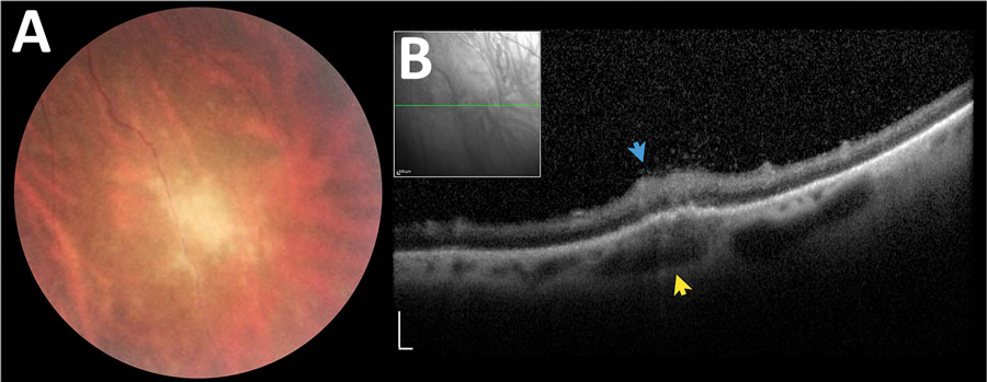
Figure 5. Asymptomatic late retinochoroiditis in right eye detected in follow-up examination at month 37 (visual acuity 0.0) in 28-year-old woman from a presumed waterborne toxoplasmosis outbreak, Brazil. A) Fundus photograph showing focal retinal whitening with indistinct borders. B) Spectral-domain optical coherence tomography showing hyper-reflectivity, disorganization, and thickening of inner retinal layers (blue arrow), and numerous overlying hyper-reflective dots at the overlying vitreous and fusiform thickening of underlying choroid (yellow arrows). Scale bars indicate 200 µm.
1Presented in part at the 2015 American Uveitis Society Fall meeting, November 15, 2014, Las Vegas, Nevada, USA
Page created: October 05, 2020
Page updated: November 19, 2020
Page reviewed: November 19, 2020
The conclusions, findings, and opinions expressed by authors contributing to this journal do not necessarily reflect the official position of the U.S. Department of Health and Human Services, the Public Health Service, the Centers for Disease Control and Prevention, or the authors' affiliated institutions. Use of trade names is for identification only and does not imply endorsement by any of the groups named above.