Volume 26, Number 6—June 2020
Research
Radical Change in Zoonotic Abilities of Atypical BSE Prion Strains as Evidenced by Crossing of Sheep Species Barrier in Transgenic Mice
Figure 1
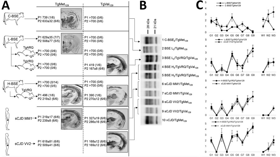
Figure 1. Atypical BSE transmission into human-PrP transgenic mice before and after adaptation to sheep PrP sequence in a study of atypical BSE transmission using isolates from different countries in Europe and transgenic mouse models overexpressing human normal brain prion protein. A) Transmission data including mean survival time + SD as well as attack rates (diseased PrPres positive/inoculated animals) and PET blot images for all atypical BSE transmission into the human-PrP transgenic mouse models. L-BSE/TgMet129 showed fine staining, and deposits were restricted to the several thalamus nuclei. C-BSE/TgMet129 showed granular deposits. L-BSE/TgVRQ/TgVal129 and TgVal129 PET blotting showed strong deposition in a particular area of the isocortex, thalamus, and midbrain, and mild deposition in the fiber tracts. H-BSE/TgVRQ and sCJD MM1 PET blotting images showed strong deposition in the isocortex area, hippocampus, thalamus, and midbrain in TgMet129 and strong deposition in the isocortex area, thalamus, and midbrain in TgVal129. B) Brain PrPres profile in TgMet129 and TgVal129 mice inoculated with atypical BSE prions before or after adaptation to the sheep-PrP sequence immunoblotted with the Sha31 mAb. Human vCJD and different sCJD prion strains inoculated in the same TgMet129 and TgVal129 mouse models are also included for comparison purposes. L-BSE/TgVRQ/TgVal129 (lane 3) is very similar to sCJD VV2/TgVal129 (lane 9). By contrast, H-BSE/TgVRQ/TgMet129 (lane 5) and sCJD MM1/TgMet129 (lane 6) are undistinguishable, as also observed with H-BSE/TgVRQ/TgVal129 (lane 4) and sCJD MM1/TgVal129 (lane 7). All PrPres profiles are different from those of vCJD/TgMet129 (lanes 1 and 10) and L-BSE/TgMet129 (lane 2). All inoculated animals were analyzed, and individual variations in the PrPres profile among them were not found. C) Vacuolar lesion profile in brains from human-PrP transgenic mice inoculated with C-BSE or the atypical BSE isolates before and after adaptation to the sheep-PrP sequence. Lesion scoring was conducted for 9 areas of gray matter (G) and 3 areas of white matter (W) in mouse brains: G1, dorsal medulla; G2, cerebellar cortex; G3, superior colliculus; G4, hypothalamus; G5, medial thalamus; G6, hippocampus; G7, septum; G8, medial cerebral cortex at the level of the thalamus; G9, medial cerebral cortex at the level of the septum (G9); W1, cerebellum; W2, mesencephalic tegmentum; W3, pyramidal tract. BSE, bovine spongiform encephalopathy; C-BSE, classical bovine spongiform encephalopathy; mAb, monoclonal antibody; PET, paraffin embedded tissue; PrP, prion protein; PrPres, proteinase K–resistant prion protein; sCJD, sporadic Creutzfeldt-Jakob disease; vCJD, variant Creutzfeldt-Jakob disease.
1These first authors contributed equally to this article.
2Current affiliation: University of California–San Diego, La Jolla, California, USA.