Volume 27, Number 4—April 2021
Research
Histopathological Characterization of Cases of Spontaneous Fatal Feline Severe Fever with Thrombocytopenia Syndrome, Japan
Figure 2
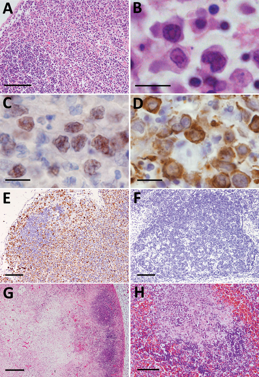
Figure 2. Histopathological lesions in the lymphoid organs from fatal cases of severe fever with thrombocytopenia syndrome (SFTS) in cats, Japan. A) Hematoxylin & eosin (HE)–stained lymph node demonstrating accumulation of blastic lymphocytes around the lymphoid follicle. Scale bar indicates 100 μm. B) HE-stained blastic lymphocytes from the lymph nodes demonstrating highly pleomorphic cells with large clear nuclei and prominent nucleoli, resembling immunoblasts. Scale bar indicates 10 μm. C, D) CD79a-stained (C) and immunohistochemistry-stained (D) blastic lymphocytes from the lymph nodes. Scale bar indicates 10 μm. E) Lymph node stained by immunohistochemistry revealing SFTS virus–positive blastic lymphocytes distributed around the follicle. Scale ar indicates 100 μm. F) Immunohistochemistry-stained hyperplastic lymph node demonstrating no SFTSV-positive cells or necrotic foci. Scale bar indicates 100 μm. G) Necrotic lymphadenitis in HE-stained lymph node. Scale bar indicates 200 μm. H) HE-stained spleen demonstrating necrotic lesions in the splenic follicle. Scale bar indicates 50 μm.
1Current affiliation: National Institute of Infectious Diseases, Tokyo, Japan.
2Current affiliation: Okayama University of Science, Ehime, Japan.