Volume 27, Number 4—April 2021
Research
Histopathological Characterization of Cases of Spontaneous Fatal Feline Severe Fever with Thrombocytopenia Syndrome, Japan
Figure 3

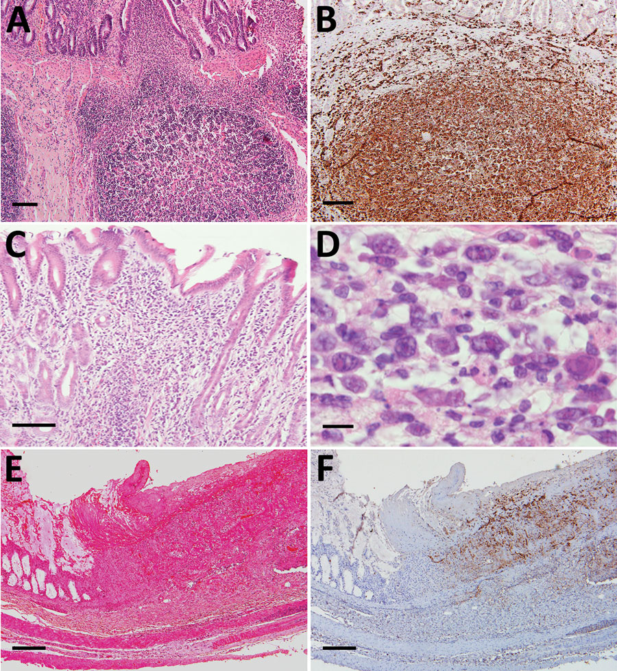
Figure 3. Histopathological lesions in the intestinal tracts from fatal cases of severe fever with thrombocytopenia syndrome (SFTS) in cats, Japan. A, B) Hematoxylin & eosin (HE)–stained (B) and immunohistochemistry-stained (A) ileum sections demonstrating enlargement of Peyer’s patch and accumulation of SFTSV-positive blastic lymphocytes. Scale bars indicates 100 μm. C) HE-stained colon sections demonstrating infiltration of lymphocytes into the lamina propria. Scale bar indicates 100 μm. D) High power magnification of panel C demonstrating the infiltrating lymphocytes were blastic lymphocytes. Scale bar indicates 10 μm. E, F) HE stained (E) and immunohistochemistry-stained (F) ulcerative lesions in the cecum. Scale bars indicate 200 μm.
1Current affiliation: National Institute of Infectious Diseases, Tokyo, Japan.
2Current affiliation: Okayama University of Science, Ehime, Japan.