Volume 27, Number 8—August 2021
Research
Modeling Immune Evasion and Vaccine Limitations by Targeted Nasopharyngeal Bordetella pertussis Inoculation in Mice
Figure 7
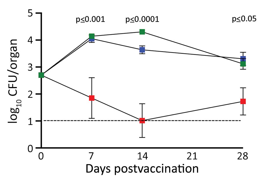
Figure 7. Comparison of nasal cavity colonization of Bordetella pertussis among experimentally infected mice after intraperitoneal vaccination with acellular pertussis (aP) or whole-cell pertussis (wP) vaccine. Graph compares colonization profiles over 28 days. Green squares indicate naive mice; blue squares indicate mice vaccinated with aP; red squares indicate mice vaccinated wP. Error bars indicate SD of the mean for 4 biologic replicates. The study was conducted twice; results are shown for a single experiment. Dotted line indicates limit of detection. p values indicate statistically significant differences between aP- and wP-vaccinated mice.
1These authors contributed equally to this article.
2Current affiliation: University of British Columbia, Vancouver, British Columbia, Canada.
3Current affiliation: Friedrich Alexander University Erlangen-Nuremberg, Erlangen, Germany.
4Current affiliation: Louisiana State University Health Science Center, Shreveport, Louisiana, USA.
5Current affiliation: Augusta University, Augusta, Georgia, USA.