Volume 28, Number 2—February 2022
Research Letter
Dirofilaria immitis Pulmonary Dirofilariasis, Slovakia
Martina Miterpáková, Daniela Antolová , Jana Rampalová, Miroslava Undesser, Tomáš Krajčovič, and Bronislava Víchová
Figure
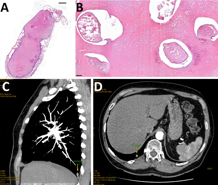
Figure. Histologic examination of resected tissue from a 66-year-old woman from southwestern Slovakia. A, B) Cross section showing Dirofilaria immitis nematodes embedded in necrotic material obtained from well-defined pulmonary nodule. Hematoxylin and eosin staining; original magnification ×20 for panel A, ×100 for panel. C, D) Chest computed tomography scan showing a subpleural focal lesion in the S10 segment of the right lung (arrows).
Page created: December 03, 2021
Page updated: January 23, 2022
Page reviewed: January 23, 2022
The conclusions, findings, and opinions expressed by authors contributing to this journal do not necessarily reflect the official position of the U.S. Department of Health and Human Services, the Public Health Service, the Centers for Disease Control and Prevention, or the authors' affiliated institutions. Use of trade names is for identification only and does not imply endorsement by any of the groups named above.