Virulence of Burkholderia pseudomallei ATS2021 Unintentionally Imported to United States in Aromatherapy Spray
Christopher K. Cote

, Kevin D. Mlynek, Christopher P. Klimko, Sergei S. Biryukov, Sherry Mou, Melissa Hunter, Nathaniel O. Rill, Jennifer L. Dankmeyer, Jeremy A. Miller, Yuli Talyansky, Michael L. Davies, J. Matthew Meinig, Stephanie A. Halasohoris, Annette M. Gray, Jade L. Spencer, Ashley L. Babyak, M. Kelly Hourihan, Bobby J. Curry, Ronald G. Toothman, Sara I. Ruiz, Xiankun Zeng, Keersten M. Ricks, Tamara L. Clements, Christina E. Douglas, Suma Ravulapalli, Christopher P. Stefan, Charles J. Shoemaker, Mindy G. Elrod, Jay E. Gee, Zachary P. Weiner, Ju Qiu, Joel A. Bozue, Nancy A. Twenhafel, and David DeShazer
Author affiliations: United States Army Medical Research Institute of Infectious Diseases, Fort Detrick, Frederick, Maryland, USA (C.K. Cote, K.D. Mlynek, C.P. Klimko, S.S. Biryukov, S. Mou, M. Hunter, N.O. Rill, J.L. Dankmeyer, J.A. Miller, Y. Talyansky, M.L. Davies, J.M. Meinig, S.A. Halasohoris, A.M. Gray, J.L. Spencer, A.L. Babyak, M.K. Hourihan, B.J. Curry, R.G. Toothman, S.I. Ruiz, X. Zeng, K.M. Ricks, T.L. Clements, C.E. Douglas, S. Ravulapalli, C.P. Stefan, C.J. Shoemaker, J. Qiu, J.A. Bozue, N.A. Twenhafel, D. DeShazer); Centers for Disease Control and Prevention, Atlanta, Georgia, USA (M.G. Elrod, J.E. Gee, Z.P. Weiner)
Main Article
Figure 9
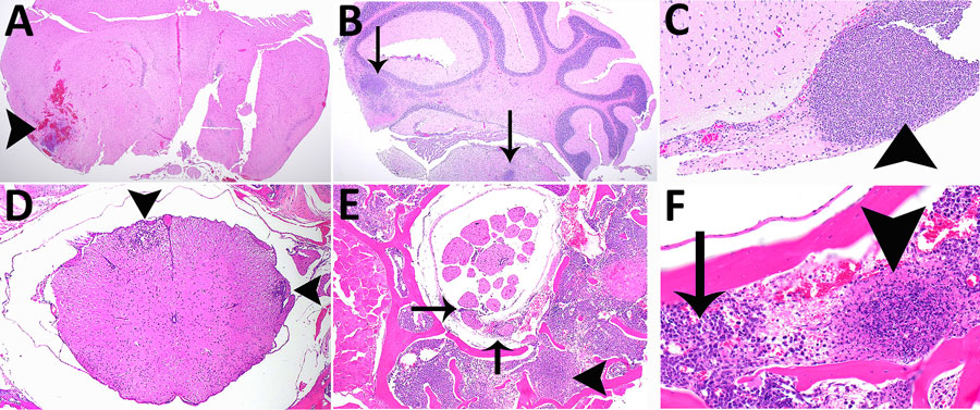
Figure 9. Histopathologic analyses of the neurologic system of C57BL/6 mice after inhalation of Burkholderia pseudomallei strain ATS2021, the causative strain in an outbreak of 4 cases, 2 of them fatal, in the United States in 2021. A) Day 5 after exposure, dose 1,150 CFU. Cerebrum showing focally extensive necrotizing and hemorrhagic meningoencephalitis (arrowhead). Hematoxylin and eosin (HE) stain; original magnification ×2. B) Day 9 after exposure, dose 107 CFU. Pons and cerebellum. There is multifocal necrotizing meningoencephalitis (arrows). HE stain; original magnification ×2. C) Day 10 after exposure, dose 1,150 CFU. Cerebrum with olfactory peduncle filled with viable and degenerate neutrophils with no recognizable peduncular tissue (arrowhead). HE stain; original magnification ×20. D) Day 9 after exposure, 107 CFU. Spinal cord, thoracic, shows multifocal meningomyelitis (arrowheads). HE stain; original magnification ×4. E) Day 5 after exposure, 1,150 CFU. Spinal cord and vertebrae, lumbar at cauda equina, show is multifocal suppurative perineuritis of spinal nerves (arrows). Note the necrotizing lesion within the vertebral bone marrow (arrowhead) HE stain; original magnification ×10. F) Day 4 after exposure, dose 1,150 CFU. Spinal cord and vertebra, thoracic, show necrotizing osteomyelitis (arrowhead). Note the loss of distinction of bone marrow cells compared to normal cells of the bone marrow (arrow). HE stain; original magnification ×40.
Main Article
Page created: September 11, 2024
Page updated: September 23, 2024
Page reviewed: September 23, 2024
The conclusions, findings, and opinions expressed by authors contributing to this journal do not necessarily reflect the official position of the U.S. Department of Health and Human Services, the Public Health Service, the Centers for Disease Control and Prevention, or the authors' affiliated institutions. Use of trade names is for identification only and does not imply endorsement by any of the groups named above.