Volume 30, Number 12—December 2024
Research
Novel Mastadenovirus Infection as Cause of Pneumonia in Imported Black-and-White Colobuses (Colobus guereza), Thailand
Figure 11
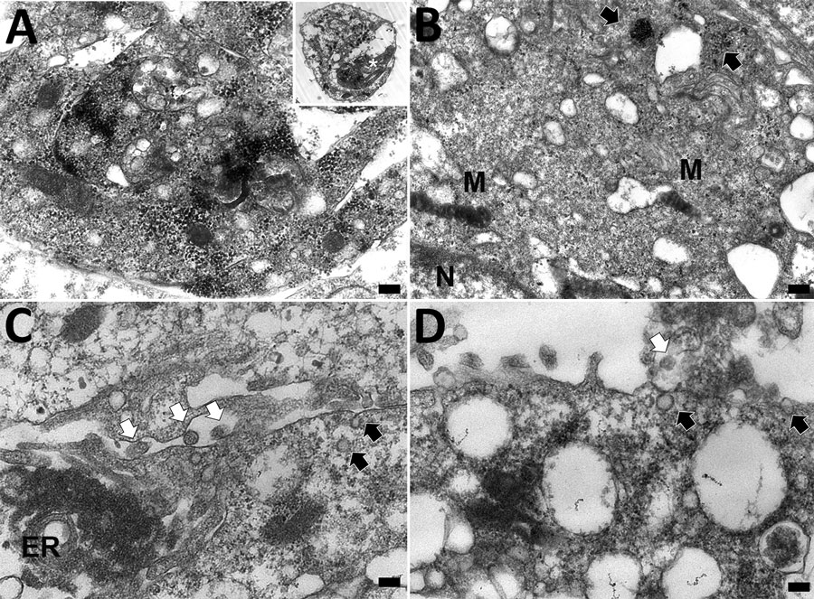
Figure 11. Transmission electron microscopy of Vero cell of case 1 in an investigation of novel mastadenovirus infection causing pneumonia in imported black-and-white colobuses (Colobus guereza), Thailand. A) Vero cells 1 day postinfection; B–C) Vero cells at 5 days postinfection A) Cytoplasmic vacuolated Vero cell contains numerous electron-dense particles packed within a destructed nucleus. Inset indicates the overall cellular morphology of an infected cell, and asterisk within indicates the area of higher magnification. Scale bar indicates 400 nm. B) Viral particles arranged within the cytosol as freely-distributed and clustered patterns (black arrows). M indicates mitochondria; N indicates nucleus. Scale bar indicates 300 nm. C) Electron-lucent viral particles within nuclear membrane and fused nuclear membrane (black arrows); electron-dense particles (white arrows) are seen within the endoplasmic reticulum (ER). Scale bar indicates 100 nm. D) A viral particle fused with plasma membrane (black arrow) and the budding virions (white arrow) in extracellular space. Scale bar indicates 100 nm.