Volume 30, Number 12—December 2024
Research
Novel Mastadenovirus Infection as Cause of Pneumonia in Imported Black-and-White Colobuses (Colobus guereza), Thailand
Figure 8
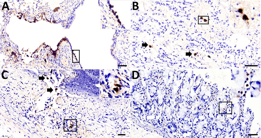
Figure 8. Adenoviral immunohistochemically stained lung and intestine sections from case 1 in an investigation novel mastadenovirus infection causing pneumonia in imported black-and-white colobuses (Colobus guereza), Thailand. A–C) Lung sections; D) intestine section. A) Nuclear labeling of adenoviral antigen in the bronchial epithelial lining; inset shows area of interest at 400× magnification. B) Adenoviral antigen is localized in the nuclei of pulmonary epithelial cells (arrows); inset shows endothelial-like cells from area of interest at 400× magnification. C) Infiltrated inflammatory cells and bronchial glands of the trachea (arrows); inset shows inflammatory cells at 400× magnification. D) Adenoviral antigen detected in rare, single inflammatory cells infiltrating the intestinal villi; inset shows area of interest at 400× magnification. Scale bars indicate 50 µm.