Volume 30, Number 7—July 2024
Dispatch
Body Louse Pathogen Surveillance among Persons Experiencing Homelessness, Canada, 2020–2021
Cite This Article
Citation for Media
Abstract
We analyzed body lice collected from persons experiencing homelessness in Winnipeg, Manitoba, Canada, during 2020–2021 to confirm vector species and ecotype and to identify louseborne pathogens. Of 556 lice analyzed from 7 persons, 17 louse pools (218 lice) from 1 person were positive for the louseborne bacterium Bartonella quintana.
In 2020, Canada’s largest cluster of Bartonella quintana endocarditis, an infection caused by a louseborne bacterium, was detected among persons experiencing homelessness in Winnipeg, Manitoba, Canada (1). Over a 6-month period, 4 people required hospitalization for B. quintana endocarditis (1). The outbreak triggered a retrospective analysis revealing 11 cases of B. quintana in Manitoba in the preceding decade (2). In 2022, the first pediatric case of B. quintana endocarditis acquired in a high-income country was reported from Manitoba (3). Prior to the Manitoba outbreak, only 3 cases of B. quintana infection were detected in Canada (4).
B. quintana is a fastidious gram-negative bacillus transmitted through the feces of infected body lice, Pediculus humanus humanus (5). The bacterium was first detected during World War I as the cause of trench fever and was later determined to cause bacteremia, endocarditis, and bacillary angiomatosis (5). B. quintana enters the bloodstream through broken skin (5).
Body lice and head lice are morphotypes of a single species, Pediculus humanus (6). Unlike head lice, body lice live in clothing, intermittently moving to the skin to feed on blood (5). Body lice are traditionally known to transmit 3 pathogens: B. quintana, Rickettsia prowazekii (epidemic typhus), and Borrelia recurrentis (louseborne relapsing fever) (5). Whereas they are not typically louseborne, Coxiella burnetii and Acinetobacter spp. have been detected in body lice (7). Body louse infestation is associated with poverty, experiencing homelessness, and an inability to wash and change clothing.
The possibility that body lice–infested persons from Winnipeg could be exposed to louseborne pathogens is unknown. In this article, we discuss what louseborne pathogens were found in Winnipeg body lice and the difference in pathogen real-time PCR cycle threshold (Ct) values according to louse instar and sex. This study was approved by the University of Manitoba and multiple other institutional ethics review boards (Appendix).
We collected ectoparasites from the clothing of participants in inner city Winnipeg. We separated ectoparasites from the same person into pools based on instar and sex. We pooled ectoparasites from the first and second instars but tested those from the third and fourth instars separately. We tested ectoparasites positive for B. quintana from the fourth instar in separate pools of male and female parasites. We decontaminated ectoparasite pools by using 70% ethanol and homogenized them by using a copper clad bead beater. We then extracted DNA by using the DNeasy 96 kit (QIAGEN, https://www.qiagen.com). We identified vector species, louse morphotype, and pathogens by using real-time PCR (Appendix). We used cytochrome b genes to identify louse species and Phum_PHUM540560 genes to identify ecotype (8). We identified pathogens by using the following targets: ITS3, Bartonella genus; yopP and fabB, B. quintana; ompB, Rickettsia prowazekii; IS1111a, Coxiella burnetii; and rpoB, Acinetobacter spp. We conducted statistical analysis by using Mann-Whitney U and Kruskal-Wallis tests (Bonferroni correction, post-hoc Dunn test) to compare groups of Ct values (Appendix). We considered values of p<0.05 significant.
Figure 1
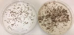
Figure 1. Body lice collected from a person experiencing homelessness in inner city Winnipeg, Manitoba, Canada. Not all ectoparasites from this person were analyzed.
Figure 2
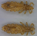
Seven persons submitted ectoparasites, 2 in 2020, and 5 in 2021 (Appendix). We analyzed 556 ectoparasites. The range of ectoparasites tested per participant was 5–218 and per pool was 5–48. We confirmed all ectoparasite pools were P. humanus humanus lice by using PCR positivity on louse and body lice targets and morphology (9) (Figures 1, 2). All louse pools from 1 participant (1/7 = 14%, 218 lice) demonstrated positivity on all Bartonella and B. quintana targets (Table 1). Of the 7 louse pools positive for B. quintana, 4 also demonstrated molecular positivity for Acinetobacter spp. Ectoparasites from all participants were negative for R. prowazekii and C. burnetii.
When analyzing B. quintana–positive louse pools, we found Ct values were similar between ITS3, yopP, and fabB genes (test statistic H = 0.54; p = 0.76). The average ITS3 Ct values decreased from the first and second instar pools (34.6) to the third instar pools (28.9) by 5.7, and from the third instar pools to the fourth instar pool (21.8) by 7.1. Pools from female lice demonstrated lower ITS3 Ct values than male lice pools (p = 0.0214) (Table 2).
We determined by molecular testing that body lice collected from a person experiencing homelessness in Winnipeg were positive for B. quintana bacteria. This finding complements the recent Manitoba cluster of B. quintana cases, suggesting a poorly described burden of infection (1,2,4). The hidden presence of B. quintana bacteria in Canada was recently highlighted in an outbreak of transplant derived B. quintana infection in cities that had not previously reported transmission: 5 cases of bacillary angiomatosis were linked to 3 deceased donors from 2 cities in Alberta (Health Canada, pers. comm., email, 2023 Nov 4). All cases were confirmed to be B. quintana bacteria with donors experiencing homelessness as the common risk factor (Health Canada, pers. comm., email, 2023 Nov 4).
Our study suggests a minority of body lice cases from Winnipeg are positive for pathogens, including B. quintana bacteria. We did not collect epidemiologic data for this study, but all participants were persons who experienced homelessness in inner city Winnipeg. Because of Winnipeg’s harsh winters and few homeless shelters, it is possible the participant with B. quintana–positive lice lives in close proximity to others and other persons with B. quintana infection remain undocumented. Only 1/7 persons with body lice had B. quintana–positive lice, which may be because of the small number of participants and that 3 participants submitted few ectoparasites. Nationwide body lice studies to compare B. quintana bacterial prevalence across different areas are needed to identify locations of infection.
The absence of other pathogens likely reflects differences in transmission dynamics and ecology (10,11). Unlike B. quintana bacteria, which does not alter louse survival, lice infected with R. prowazekii bacteria die within a week of infection, limiting transmission (11). The urban setting of our study diminishes the chance of replicating the occasional documentation of C. burnetii bacteria in lice. Whereas Acinetobacter spp. bacteria are commonly identified in body lice, no proven cases of Acinetobacter disease caused by body lice have been confirmed (11,12).
The lower Bartonella Ct values (stronger signal) with advancing louse instar and female sex may indicate larger blood meals of those subpopulations. B. quintana bacteria replicate in the louse intestine but are not known to be transmitted transovarially, indicating the person with B. quintana–positive lice from all instars likely had sustained bacteremia for at least 1 month (body lice lifespan). This study highlights the usefulness of identifying ectoparasites by using molecular methods when arthropod taxonomic expertise is not accessible.
B. quintana bacteria is excreted in louse feces continuously for weeks in quantities up to 107 bacteria/louse each day (13,14). The explosive replication, coupled with B. quintana bacteria remaining infectious in biofilm-like structures for up to 1 year, suggests even a single case of B. quintana infection may indicate a hidden burden of infected persons (5,14).
Our study is limited by a small sample size, the heterogenous number of ectoparasites submitted per person, the focus on urban populations from 1 jurisdiction, and the lack of DNA quantity normalization. Active case finding, contact tracing, and public health engagement are needed to clarify the epidemiology of B. quintana infection in Canada. Manitoba residents with body lice should be evaluated for B. quintana infection. Sampling of ectoparasites may provide an effective way to perform surveillance for emerging pathogens in marginalized settings.
Dr. Boodman is an infectious disease doctor and medical microbiologist who is currently a PhD candidate at the Institute of Tropical Medicine (Belgium) and the University of Antwerp (Belgium), supported by University of Manitoba’s Clinical Investigator Program (Canada). His interests include neglected infections linked to poverty and vectorborne intracellular bacteria.
References
- Boodman C, Wuerz T, Lagacé-Wiens P. Endocarditis due to Bartonella quintana, the etiological agent of trench fever. CMAJ. 2020;192:E1723–6. DOIPubMedGoogle Scholar
- Boodman C, Wuerz T, Lagacé-Wiens P, Lindsay R, Dibernardo A, Bullard J, et al. Serologic testing for Bartonella in Manitoba, Canada, 2010–2020: a retrospective case series. CMAJ Open. 2022;10:E476 LP–E482.
- Boodman C, MacDougall W, Hawkes M, Tyrrell G, Fanella S. Bartonella quintana endocarditis in a child from Northern Manitoba, Canada. PLoS Negl Trop Dis. 2022;16:
e0010399 . DOIPubMedGoogle Scholar - Lam JC, Fonseca K, Pabbaraju K, Meatherall BL. Case report: Bartonella quintana endocarditis outside of the Europe-African gradient: comprehensive review of cases within North America. Am J Trop Med Hyg. 2019;100:1125–9. DOIPubMedGoogle Scholar
- Foucault C, Brouqui P, Raoult D. Bartonella quintana characteristics and clinical management. Emerg Infect Dis. 2006;12:217–23. DOIPubMedGoogle Scholar
- Light JE, Toups MA, Reed DL. What’s in a name: the taxonomic status of human head and body lice. Mol Phylogenet Evol. 2008;47:1203–16. DOIPubMedGoogle Scholar
- Amanzougaghene N, Fenollar F, Raoult D, Mediannikov O. Where are we with human lice? A review of the current state of knowledge. Front Cell Infect Microbiol. 2020;9:474. DOIPubMedGoogle Scholar
- Drali R, Boutellis A, Raoult D, Rolain JM, Brouqui P. Distinguishing body lice from head lice by multiplex real-time PCR analysis of the Phum_PHUM540560 gene. PLoS One. 2013;8:
e58088 . DOIPubMedGoogle Scholar - Bonilla DL, Durden LA, Eremeeva ME, Dasch GA. The biology and taxonomy of head and body lice—implications for louse-borne disease prevention. PLoS Pathog. 2013;9:
e1003724 . DOIPubMedGoogle Scholar - Bonilla DL, Kabeya H, Henn J, Kramer VL, Kosoy MY. Bartonella quintana in body lice and head lice from homeless persons, San Francisco, California, USA. Emerg Infect Dis. 2009;15:912–5. DOIPubMedGoogle Scholar
- Badiaga S, Raoult D, Brouqui P. Preventing and controlling emerging and reemerging transmissible diseases in the homeless. Emerg Infect Dis. 2008;14:1353–9. DOIPubMedGoogle Scholar
- La Scola B, Raoult D. Acinetobacter baumannii in human body louse. Emerg Infect Dis. 2004;10:1671–3. DOIPubMedGoogle Scholar
- Chomel BB, Boulouis H-J, Breitschwerdt EB, Kasten RW, Vayssier-Taussat M, Birtles RJ, et al. Ecological fitness and strategies of adaptation of Bartonella species to their hosts and vectors. Vet Res. 2009;40:29. DOIPubMedGoogle Scholar
- Seki N, Kasai S, Saito N, Komagata O, Mihara M, Sasaki T, et al. Quantitative analysis of proliferation and excretion of Bartonella quintana in body lice, Pediculus humanus L. Am J Trop Med Hyg. 2007;77:562–6. DOIPubMedGoogle Scholar
Figures
Tables
Cite This ArticleTable of Contents – Volume 30, Number 7—July 2024
| EID Search Options |
|---|
|
|
|
|
|
|
Please use the form below to submit correspondence to the authors or contact them at the following address:
Carl Boodman, Max Rady College of Medicine, University of Manitoba, Rm 543 Basic Medical Sciences Bdg, 745 Bannatyne Ave, Winnipeg, MN R3E 0J9, Canada
Top