Volume 30, Number 7—July 2024
Dispatch
Avian Influenza A(H5N1) Virus among Dairy Cattle, Texas, USA
Cite This Article
Citation for Media
Abstract
During March and April 2024, we studied dairy cattle specimens from a single farm in Texas, USA, using multiple molecular, cell culture, and next-generation sequencing pathogen detection techniques. Here, we report evidence that highly pathogenic avian influenza A(H5N1) virus strains of clade 2.3.4.4b were the sole cause of this epizootic.
Since the arrival of clade 2.3.4.4b avian influenza A(H5N1) in North America in late 2021, frequent mammal spillover events have occurred in a diverse range of species, including 1 human infection, but those strains have not affected cattle. Cattle are known to be permissive but resilient to infection with influenza A, B, and C viruses (1); however, they are susceptible to influenza D virus, which is thought to have near-worldwide distribution (2). Influenza D virus is thought to move from cow-to-cow through direct contact or short-distance aerosol respiratory transmission (2), and possible occasional influenza D virus spillover to humans is a concern (3,4). Even so, influenza viruses are not the first pathogens veterinarians or veterinary diagnostic laboratories search for in studying cattle respiratory epizootics. We report results of an investigation into influenza virus infections among dairy cattle on a farm in Texas, USA.
On March 18, 2024, we were notified of epidemics of illness among Texas dairy cattle. The cattle had transient respiratory and gastrointestinal signs (5). Veterinary diagnostic laboratory results were largely unremarkable except for rumors among cattle veterinarians of possible influenza A virus detection among cattle and conjunctivitis among dairy farm workers. The University of Texas Medical Branch (UTMB) research team offered diagnostic support owing to the team’s novel pathogen detection capabilities (6–9) and having recognized that conjunctivitis among workers handling animals had been previously noted in association with highly pathogenic avian influenza (HPAI) epizootics (10–13). On March 19, we were invited to investigate the outbreak by a farm owner. We provided the farm with sampling supplies and instructions. UTMB’s Institutional Animal Care and Use Committee has viewed such diagnostic work to be exempt from formal ethics review.
To determine the etiology of cattle illnesses, we used molecular screening and, in some cases, cell culture and metagenomics, to examine cattle swab specimens (Appendix 1). We targeted 6 viral groups, adenoviruses, coronaviruses, enteroviruses, influenza viruses, paramyxoviruses, and pneumoviruses, using previously published techniques (8).
At our request, on March 21, dairy farm management collected and shipped swab specimens from the nasal passages of 14 cows with signs of illness and 6 cows with no sign of illness in a shipping container with ice packs. We received the samples and completed questionnaires on March 22. To determine whether pathogens were enteric, we requested additional samples from the dairy farm on March 28. Nasal and rectal swab specimens were taken from 10 additional ill cows on April 1; we received those 20 additional swab specimens on April 3.
The 40 swab specimens were obtained from 30 different cows (24 sick and 6 healthy) from the same dairy farm (Appendix 1 Table 1); specific farm location, name, and cattle breed are withheld for privacy purposes. Sampled cattle ranged from 2 years 3 months of age to 7 years 10 months of age.
Farm staff first observed illnesses in cattle on March 6. Cattle with otherwise healthy records showed signs of decreased appetite, lethargy, increased respiratory secretions, high temperatures (up to 105°F or 40.56°C), abnormal bowel movements, and decreased milk production. During March 10–12, >4.75% of the herd had clinical signs of influenza-like illness and were being treated in the hospital pens. No dead birds, dead cats, or other deceased wildlife were observed. At the time of specimen collections, cattle illnesses were on the wane.
Several workers experienced influenza-like symptoms and missed work during March 4–6. A maternity worker visited a local clinic and received treatment for influenza-like symptoms; 2 milkers also experienced influenza-like symptoms and stayed home. No cases of conjunctivitis, severe illness, or hospitalizations were reported among workers.
Figure 1
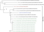
Figure 1. Phylogenetic tree of the concatenated genome in study of avian influenza A(H5N1) virus among dairy cattle, Texas, USA. Maximum-likelihood phylogenetic tree inferred for the A/cattle/Texas/5628356283/2024 (H5N1) virus isolated in this...
Figure 2
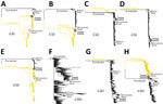
Figure 2. Phylogenetic trees for 8 genome segments in study of avian influenza A(H5N1) virus among dairy cattle, Texas, USA. Maximum-likelihood phylogenetic trees inferred for each of the 8 segments of the...
Multiple cattle swab specimens demonstrated molecular evidence of H5 avian influenza A virus (Appendix 1 Table 1). Of the first 20 cattle swab samples received, none had evidence of adenovirus, coronavirus, enterovirus, or influenza D. Of those first 20 specimens, 3 (2 healthy cows, 1 sick cow) demonstrated molecular evidence of a Paramyxoviridae or Pneumoviridae virus (Appendix 1 Table 1). Multiple swab cultures in MDBK, Vero E6, and MDCK cells also had molecular evidence of H5 avian influenza A virus. Molecular study of the HA cleavage site demonstrated that those viruses were highly pathogenic. Next-generation sequencing (NGS) corroborated these findings; 1 cultured cattle nasal swab specimen yielded a complete genome A/cattle/Texas/56283/2024 (H5N1) (GenBank accession nos. PP600140–7 for the 8 viral segments), confirmed to be HPAI and of clade 2.3.4.4b. We performed phylogenetic comparisons of related viruses in GenBank and GISAID (https://www.gisaid.org) for the entire genome (Figure 1) and the virus’s 8 gene segments (Figure 2), which documented similarity to 13 other viruses in the Texas epizootic clade. A/cattle/Texas/56283/2024 (H5N1) had several novel mutations in comparison to related viruses (Table). One mutation (PB2-M631L) increases the capability of H5N1 to replicate in human cells by enhancing the polymerase activity of the viruses in human cells. Pathogenicity studies in animal models will be necessary to better understand such viruses. NGS analyses suggested that the sick cow in which a nasal swab specimen tested positive for Paramyxoviridae or Pneumoviridae virus (cattle identification [ID] 49869) (Appendix 1 Table 1) had a bovine viral diarrhea virus (BVDV), indicating a possible cause of illness.
This preliminary study of a single Texas dairy farm affected by what is now a multistate epizootic of HPAI H5N1 documents several key observations. H5N1 virus detections were made solely in the sick cows without apparent co-infecting viruses (5 other viral families examined). HPAI H5N1 virus was more prevalent among nasal swab samples than rectal swab samples, supporting the notion that the respiratory tract of cattle could be involved in cow-to-cow transmission.
Although 1 sick cow (cattle ID 49869; Appendix 1 Table 1) was found by NGS to have evidence of a BVDV in its nasal swab specimen, 2 other healthy cows also had panspecies evidence of such a virus (cattle IDs 74061 and 54972; Appendix 1 Table 1); however, we did not perform NGS on their specimens. BVDVs are frequently associated with mild respiratory disease on cattle farms; because this farm routinely administers a vaccine with live BVDV components, we doubt that the BVDV explains the unusual illness seen in this farm’s dairy cattle herd.
The complete genome of the H5N1 virus isolated from 1 sick cow’s nasal swab specimen suggests that this H5N1 strain is very similar to the H5N1 strains characterized from dead birds, other cattle (14), and 1 cattle worker (15). The high genetic similarity of A/cattle/Texas/56283/2024 (H5N1) and other avian, human, and cattle strains in the Texas clade suggests a single interconnected multispecies outbreak in Texas, the precise directions of transmission still to be determined.
A limitation of our study is that we only examined specimens sent to us. We did not collect milk, study animal workers, or collect environmental specimens, nor did we immediately visit the farm for a comprehensive outbreak investigation. However, many barriers to performing a more traditional outbreak investigation on HPAI-infected farms currently exist.
The ongoing multispecies HPAI H5N1 outbreak involving birds, cattle, goats, alpacas, humans, cats, and other species epitomizes why interdisciplinary cooperation under a One Health framework is required. If we wish to resolve complex problems such as this epizootic, finding ways to assure farm owners that necessary epidemiological investigations will not harm their businesses will be imperative.
Dr. Oguzie is a veterinarian, molecular biologist, and current postdoctoral fellow at the University of Texas Medical Branch in Galveston, Texas. Her primary research interests are molecular surveillance and genomic characterization of novel pathogens. Dr. Marushchak is a veterinary scientist and postdoctoral fellow at the University of Texas Medical Branch in Galveston, Texas. Her primary research interest is conducting molecular surveillance for emerging pathogens at the human-animal interface.
Acknowledgments
We thank Diego Silva and Claudia Trijillo for their administrative and laboratory support of this work. We thank Robert H. Carpenter and Kay Russo for their education regarding livestock farming. We thank the dairy farm owners for engaging us in research collaboration. We gratefully acknowledge all data contributors (i.e., the authors and their originating laboratories responsible for obtaining the specimens and their submitting laboratories for generating the genetic sequence and metadata and sharing via the GISAID Initiative) on which this research is based (Appendix 2).
Data needed to evaluate the conclusions in the paper are present in the paper and the Appendices. Researchers with BSL3Ag-approved laboratories may request A/cattle/Texas/56283/2024 (H5N1) by contacting Kenneth Plante (
This work was supported by G.C.G.’s startup funding from the University of Texas Medical Branch and in support of M.I.N.’s work, by the Intramural Research Program at the National Library of Medicine at the National Institutes of Health and the Centers of Excellence for Influenza Research and Surveillance, National Institute of Allergy and Infectious Diseases, National Institutes of Health, Department of Health and Human Services (contract HHSN272201400006C).
G.C.G. conceptualized the study. J.U.O., L.V.M., I.S., A.L.M., J.A.L., H.H., and G.C.G. constructed the methodology. Investigation was carried out by G.C.G., J.U.O., L.V.M., I.S., A.L.M., J.A.L., and H.H. M.I.N. and J.U.O. performed data analysis. Visualization was performed by J.U.O. and H.H. Funding was acquired by G.C.G., and the project was administered by G.C.G. and L.V.M. G.C.G. and L.V.M. supervised the study. J.U.O. and G.C.G. wrote the original draft, and all authors contributed to the review and editing of the final manuscript.
References
- Sreenivasan CC, Sheng Z, Wang D, Li F. Host range, biology, and species specificity of seven-segmented influenza viruses—a comparative review on influenza C and D. Pathogens. 2021;10:1583. DOIPubMedGoogle Scholar
- Ruiz M, Puig A, Bassols M, Fraile L, Armengol R, Influenza D. Influenza D virus: a review and update of its role in bovine respiratory syndrome. Viruses. 2022;14:2717. DOIPubMedGoogle Scholar
- White SK, Ma W, McDaniel CJ, Gray GC, Lednicky JA. Serologic evidence of exposure to influenza D virus among persons with occupational contact with cattle. J Clin Virol. 2016;81:31–3. DOIPubMedGoogle Scholar
- Leibler JH, Abdelgadir A, Seidel J, White RF, Johnson WE, Reynolds SJ, et al. Influenza D virus exposure among US cattle workers: A call for surveillance. Zoonoses Public Health. 2023;70:166–70. DOIPubMedGoogle Scholar
- American Association of Bovine Practitioners. AABP statement on Texas cattle outbreak. 2024 Mar 18 [cited 2024 Mar 18]. https://agpartners.net/2024/03/18/aabp-statement-on-texas-cattle-outbreak
- Ramesh A, Bailey ES, Ahyong VLC, Phelps M, Neff N, Sit R, et al. Microbial diversity in a North American swine farm operation. Sci Rep. 2021;11:16994. DOIPubMedGoogle Scholar
- Xiu L, Binder RA, Alarja NA, Kochek K, Coleman KK, Than ST, et al. A RT-PCR assay for the detection of coronaviruses from four genera. J Clin Virol. 2020;128:
104391 . DOIPubMedGoogle Scholar - Gray GC, Robie ER, Studstill CJ, Nunn CL. Mitigating future respiratory virus pandemics: new threats and approaches to consider. Viruses. 2021;13:637. DOIPubMedGoogle Scholar
- Vlasova AN, Diaz A, Damtie D, Xiu L, Toh TH, Lee JS, et al. Novel canine coronavirus isolated from a hospitalized patient with pneumonia in east Malaysia. Clin Infect Dis. 2022;74:446–54. DOIPubMedGoogle Scholar
- Arzey GG, Kirkland PD, Arzey KE, Frost M, Maywood P, Conaty S, et al. Influenza virus A (H10N7) in chickens and poultry abattoir workers, Australia. Emerg Infect Dis. 2012;18:814–6. DOIPubMedGoogle Scholar
- Liu Q, Liu DY, Yang ZQ. Characteristics of human infection with avian influenza viruses and development of new antiviral agents. Acta Pharmacol Sin. 2013;34:1257–69. DOIPubMedGoogle Scholar
- Belser JA, Creager HM, Zeng H, Maines TR, Tumpey TM. Pathogenesis, transmissibility, and tropism of a highly pathogenic avian influenza A(H7N7) virus associated with human conjunctivitis in Italy, 2013. J Infect Dis. 2017;216(suppl_4):S508–11. DOIPubMedGoogle Scholar
- Creager HM, Kumar A, Zeng H, Maines TR, Tumpey TM, Belser JA. Infection and replication of influenza virus at the ocular surface. J Virol. 2018;92:e02192–17. DOIPubMedGoogle Scholar
- Burrough ER, Magstadt DR, Petersen B, Timmermans SJ, Gauger PC, Zhang J, et al. Highly pathogenic avian influenza A(H5N1) clade 2.3.4.4b virus infection in domestic dairy cattle and cats, United States, 2024. Emerg Infect Dis. 2024;30:30. DOIPubMedGoogle Scholar
- Uyeki TM, Milton S, Abdul Hamid C, Reinoso Webb C, Presley SM, Shetty V, et al. Highly pathogenic avian influenza A(H5N1) virus infection in a dairy farm worker. N Engl J Med. N Engl J Med. 2024;•••:
c2405371 .
Figures
Table
Cite This ArticleOriginal Publication Date: June 07, 2024
1These first authors contributed equally to this article.
Table of Contents – Volume 30, Number 7—July 2024
| EID Search Options |
|---|
|
|
|
|
|
|
Please use the form below to submit correspondence to the authors or contact them at the following address:
Gregory C. Gray, Infectious Disease Epidemiology, Departments of Internal Medicine (Infectious Diseases), Microbiology and Immunology, and Global Health, University of Texas Medical Branch, 301 University Blvd, Rte 0435, Galveston, TX 77555, USA
Top