Volume 30, Number 9—September 2024
Dispatch
Autochthonous Human Babesiosis Caused by Babesia venatorum, the Netherlands
Niekie Spoorenberg1, Clara F. Köhler1, Evelien Vermeulen1, Suzanne Jurriaans, Marion Cornelissen, Kristina E.M. Persson, Iris van Doorn, Hein Sprong, Joppe W. Hovius, and Rens Zonneveld
Figure 1
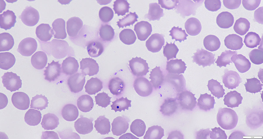
Figure 1. Babesia venatorum parasites in blood of patient with autochthonous human babesiosis, the Netherlands. Giemsa-stained thin smear from blood of patient before start of treatment and visualized with light microscopy (original magnification ×1,250) shows small and pleomorphic amoeboid and vacuolar asexual stages of Babesia spp., positioned within and outside red blood cells, which is typically observed in human babesiosis. Of note, 1 dark blue–stained Howell-Jolly body is observed.
1These authors contributed equally to this article.
Page created: August 01, 2024
Page updated: August 21, 2024
Page reviewed: August 21, 2024
The conclusions, findings, and opinions expressed by authors contributing to this journal do not necessarily reflect the official position of the U.S. Department of Health and Human Services, the Public Health Service, the Centers for Disease Control and Prevention, or the authors' affiliated institutions. Use of trade names is for identification only and does not imply endorsement by any of the groups named above.