Volume 31, Number 1—January 2025
Dispatch
Domestic Cat Hepadnavirus Infection in Iberian Lynxes
Abstract
We conducted a survey for domestic cat hepadnavirus, an analog of human hepatitis B virus, in the endangered felid species Iberian lynx. Results revealed specific antibodies in 32.3% of serum samples and DNA in 0.5% of available liver samples. Phylogenetically, the virus segregated apart from other Europe strains of the virus.
Domestic cat hepadnavirus (DCH) is a novel member of the genus Orthohepadnavirus, family Hepadnaviridae, similar to the prototype species hepatitis B virus (HBV). The virus was first documented in 2018 in Australia in a domestic cat with lymphoma; since then, the virus has been described in cats all over the world (1,2). The DCH genome is a circular, partially double-stranded DNA, ≈3.2 kb in length, containing 2 large and 2 smaller open reading frames, encoding for the surface protein, the polymerase protein, the precore/core protein, and the X protein (1).
HBV infection is a global health challenge representing a major cause of chronic liver diseases in humans, including cirrhosis and hepatocellular carcinoma (2). Similarly, reports have correlated DCH with development of feline liver disease and identified the virus in cats with chronic hepatitis and cats with hepatocellular carcinoma (2–4), stimulating research to investigate the possible implications for feline health. Researchers have reported the virus, at a very low prevalence, also in dogs (5); however, studies assessing the susceptibility of other animal hosts to DCH or DCH-like viruses remain elusive.
The Iberian lynx (Lynx pardinus) is the most endangered felid species in the world (6). By the early 21st Century, Iberian lynx population was estimated to include 156 adult animals in Portugal and Spain (6). In response to those findings, conservation organizations launched projects focusing on both in situ and ex situ conservation programs, one of which was the European Commission’s EU LIFE-Nature and Biodiversity programme (7). Because of such efforts, the Iberian lynx census has increased considerably during the past decade, reaching >1,600 free-ranging lynxes in 2022. Amid the conservation activities surrounding this species emerged investigations into the pathogens that could pose threats to these animals, such as SARS-CoV-2 and feline leukemia virus (8,9). We investigated the exposure of Iberian lynxes to DCH.
We performed a survey on liver samples collected from 191 Iberian lynxes subjected to necroscopy in 2017–2023 throughout the Iberian Peninsula. Our screening also included 103 serum samples obtained from 100 lynxes affiliated with health programs in a 14-year time frame spanning 2010–2023. Both liver and serum samples were available for 7 lynxes. We obtained all samples from serum and tissue banks at the Center for Analysis and Diagnosis of Wildlife (Andalusia, Spain) and stored them at −80°C before shipment to the Department of Veterinary Medicine, University of Bari (Bari, Italy) for the analyses. We homogenized (10% wt/vol) liver tissues in Dulbecco modified Eagle’s medium and extracted viral DNA from the supernatant of the homogenates and from the serum by using the IndiSpin Pathogen Kit (Indical Bioscience GmbH, https://www.indical.com). We screened DNA extracts for the presence of DCH by using a quantitative PCR (10) and a qualitative PCR with panhepadnavirus primers targeting the polymerase ORF (11).
Our analyses revealed 1 (0.5%) of the 191 liver samples testing positive for viral DNA by qualitative PCR; none of the serum samples contained viral DNA. We traced the DCH-positive sample to a 7-year-old male Iberian lynx, raised in captivity in Andalusia in southern Spain (collection date March 2021). Samples for the animal included 4 serum samples collected over a 5-year period, 2016–2020; screening showed DCH DNA in only the fourth serum sample (collection date December 2020). We performed DNA enrichment for the DCH-positive liver sample (SPA/2022/Iberian lynx/296-23-81 strain) by using a rolling circle amplification technique with a TempliPhi 100 amplification kit (GE Healthcare, https://www.gehealthcare.com). We used the amplification product as a template for amplifying DCH genome fragments in PCR. A total of 500 ng of equimolar pooled PCR products made up the input for a library prepared using the Ligation Sequencing Kit V14 (Oxford Nanopore Technology, https://nanoporetech.com), according to manufacturer’s guidelines. We performed sequencing by using flongle flow cell R10.4.1 adapted in the MinION Mk1C platform (Oxford Nanopore Technology) for 24 hours.
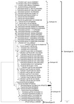
We generated the complete DCH genome of the SPA/2022/Iberian lynx/296-23-81 strain (GenBank accession no. PP347721) measuring 3,184 bp in length. The Iberian lynx strain displayed 98.3% nucleotide identity to the Thailand strain CP87H_THA/2019 (GenBank accession no. MT506044) and <96.5% nucleotide identity to other DCH strains from Europe. On phylogenetic analysis, the strain SPA/2022/Iberian lynx/296-23-81 segregated with Thailand DCH strains within genotype A, into the distinct subtype A3, apart from other DCH strains from Europe, which segregated within either subtype A1 or subtype A2 (Figure).
We tested all serum samples at a dilution of 1:100 by using 2 in-house ELISA assays, one based on the recombinant core (DCHcAg) antigen and one based on the surface (DCHsAg) antigen (12,13), to evaluate the serologic response against DCH. The 4 serum samples collected at different points from the DCH-positive Iberian lynx reacted for DCHcAg IgG but not for DCHcAg IgM or DCHsAg IgG. Our testing detected viral DNA only in the last serum sample from the animal. That pattern is consistent with the status of HBV reactivation, characterized by a peak of viremia in persons with inactive infection, wherein the virus is barely detectable in the serum although replicating in the liver (3). Our analysis also revealed DCHcAg IgG in 32 (32.3%) of 99 serum samples, with the highest detection rate in adult free-ranging lynxes (7/16, 43.8%), and no DCHcAg IgM. Only 10 (31.2%) of the 32 serum samples with DCHcAg IgG had also DCHsAg IgG, suggesting clearance from the infection. In humans, HBVcAg IgG is persistent and indicative of exposure to HBV, regardless of the evolutive stage of the infection (14).
Our study results provide evidence for a wide circulation of DCH in the Iberian lynx population, with a seroprevalence rate (32.3%) higher than those observed in cats (25%) and dogs (10%) in Italy (12,13). We propose the need for additional studies to assess the effect of this virus on the health status of the Iberian lynx. Because cats are considered the primary reservoir of feline leukemia virus infection for the lynx population (9), it will be specifically important to investigate the role of domestic cats as a potential source of DCH infection for lynxes.
DCH appears to follow a pattern similar to that of HBV, presenting different types and subtypes based on nucleotide sequence diversity. In HBV, genotypes and subgenotypes might even play a crucial role in clinical outcomes, influencing disease evolution and drug resistance (15). In our study, the Iberian lynx DCH strain did not segregate phylogenetically with other DCH strains from Europe detected in cats, raising questions as to the epidemiology of DCH and whether DCH subtype A3 exists in feline populations across the Iberian Peninsula or whether it is a hallmark of the Iberian lynx population. As more clinical and epidemiologic research on DCH unfolds, so might a greater understanding of whether different DCH types and subtypes exhibit phenotypic variations.
The viromes of closely related animal species, or even of species more distant in the evolutionary scale, are largely interconnected, with repeated events of interspecies transmissions and several examples of successful adaptation. Still, the patterns of infection and disease of viruses in a heterologous species remain unpredictable. The One Health model recommends intensifying the efforts in the study of animal pathogens to improve animal health and welfare and ensure animal conservation. This approach strongly applies to endangered animal species such as Iberian lynx.
Dr. Diakoudi is a researcher at the University of Bari, Bari, Italy. Her research interests cover virus discovery in animals, with a particular focus on viruses with zoonotic potential.
Acknowledgments
We thank all the veterinarians and animal keepers of ex situ and in situ conservation programs involved in the sampling as well as all the members of the Center for Analysis and Diagnosis of Wildlife (Spain) for their assistance in the collection of samples and epidemiological information. We also gratefully acknowledge Junta de Andalucía and Junta de Comunidades de Castilla-La Mancha.
This study did not involve the purposeful killing of animals. Samples included in this study were taken from serum banks or animals subjected to either medical check-ups, health programs, or surgical interventions during the study period. Samples from Iberian lynxes were collected by authorized veterinarians and animal keepers following routine procedures from alive or dead persons before the design of the study, in compliance with the Ethical Principles in Animal Research. Thus, ethics approval by an Institutional Animal Care and Use Committee was not deemed necessary.
This article is based upon work from project LIFE 19NAT/ES001055 LYNXCONNECT “Creating a genetically and demographically functional Iberian Lynx (Lynx pardinus) metapopulation (2020–2025),” supported by the European Commission. This research was supported by European Union funding within the MUR PNRR Extended Partnership initiative on Emerging Infectious Diseases (project no. PE00000007, INF-ACT) and by the MUR PRIN 2022 project “Investigating hepatotropic viruses in carnivores and humans in a One Health perspective” (project no. 2022EPP2TT, HVOH). This work was also supported by the National Laboratory for Infectious Animal Diseases, Antimicrobial Resistance, Veterinary Public Health and Food Chain Safety, RRF-2.3.1-21-2022-00001. Our research was also partially supported by the CIBER–Consorcio Centro de Investigación Biomédica en Red (CB 2021/13/00083), Instituto de Salud Carlos III, Ministerio de Ciencia e Innovación, and Unión Europea–NextGeneration EU. J.C.-G. was supported by the CIBER–Consorcio Centro de Investigación Biomédica en Red (CB21/13/00083), Instituto de Salud Carlos III, Ministerio de Ciencia e Innovación, and Unión Europea–Next Generation EU. S.C.-S. was supported by an FPU grant from the Spanish Ministry of Universities (FPU19/06026).
References
- Aghazadeh M, Shi M, Barrs VR, McLuckie AJ, Lindsay SA, Jameson B, et al. A novel hepadnavirus identified in an immunocompromised domestic cat in Australia. Viruses. 2018;10:269. DOIPubMedGoogle Scholar
- Shofa M, Kaneko Y, Takahashi K, Okabayashi T, Saito A. Global prevalence of domestic cat hepadnavirus: an emerging threat to cats’ health? Front Microbiol. 2022;13:
938154 . DOIPubMedGoogle Scholar - Piewbang C, Dankaona W, Poonsin P, Yostawonkul J, Lacharoje S, Sirivisoot S, et al. Domestic cat hepadnavirus associated with hepatopathy in cats: A retrospective study. J Vet Intern Med. 2022;36:1648–59. DOIPubMedGoogle Scholar
- Capozza P, Pellegrini F, Camero M, Diakoudi G, Omar AH, Salvaggiulo A, et al. Hepadnavirus infection in a cat with chronic liver disease: a multi-disciplinary diagnostic approach. Vet Sci. 2023;10:668. DOIPubMedGoogle Scholar
- Diakoudi G, Capozza P, Lanave G, Pellegrini F, Di Martino B, Elia G, et al. A novel hepadnavirus in domestic dogs. Sci Rep. 2022;12:2864. DOIPubMedGoogle Scholar
- Rodríguez A, Calzada J. The IUCN Red List of Threatened Species: Lynx pardinus. 2015 [cited 2024 Apr 23] https://www.iucnredlist.org/species/pdf/174111773
- Vargas A. Iberian Lynx ex situ conservation: an interdisciplinary approach. Vargas A, Breitenmoser-Wursten C, Breitenmoser U, editors. Fundacion Biodiversidad. 2009 [cited DATE]. https://portals.iucn.org/library/node/10167
- Gómez JC, Cano-Terriza D, Segalés J, Vergara-Alert J, Zorrilla I, Del Rey T, et al. Exposure to severe acute respiratory syndrome coronavirus 2 (SARS-CoV-2) in the endangered Iberian lynx (Lynx pardinus). Vet Microbiol. 2024;290:
110001 . DOIPubMedGoogle Scholar - Nájera F, López G, Del Rey-Wamba T, Malik RA, Garrote G, López-Parra M, et al. Long-term surveillance of the feline leukemia virus in the endangered Iberian lynx (Lynx pardinus) in Andalusia, Spain (2008-2021). Sci Rep. 2024;14:5462. DOIPubMedGoogle Scholar
- Lanave G, Capozza P, Diakoudi G, Catella C, Catucci L, Ghergo P, et al. Identification of hepadnavirus in the sera of cats. Sci Rep. 2019;9:10668. DOIPubMedGoogle Scholar
- Wang B, Yang XL, Li W, Zhu Y, Ge X-Y, Zhang L-B, et al. Detection and genome characterization of four novel bat hepadnaviruses and a hepevirus in China. Virol J. 2017;14:40. DOIPubMedGoogle Scholar
- Fruci P, Palombieri A, Sarchese V, Aste G, Friedrich KG, Martella V, et al. Serological and molecular survey on domestic dog hepadnavirus in household dogs, Italy. Animals (Basel). 2023;13:729. DOIPubMedGoogle Scholar
- Fruci P, Di Profio F, Palombieri A, Massirio I, Lanave G, Diakoudi G, et al. Detection of antibodies against domestic cat hepadnavirus using baculovirus-expressed core protein. Transbound Emerg Dis. 2022;69:2980–6. DOIPubMedGoogle Scholar
- Schillie S, Vellozzi C, Reingold A, Harris A, Haber P, Ward JW, et al. Prevention of hepatitis B virus infection in the United States: recommendations of the Advisory Committee on Immunization Practices. MMWR Recomm Rep. 2018;67:1–31. DOIPubMedGoogle Scholar
- Chen J, Li L, Yin Q, Shen T. A review of epidemiology and clinical relevance of Hepatitis B virus genotypes and subgenotypes. Clin Res Hepatol Gastroenterol. 2023;47:
102180 . DOIPubMedGoogle Scholar
Figure
Cite This ArticleOriginal Publication Date: December 11, 2024
Table of Contents – Volume 31, Number 1—January 2025
| EID Search Options |
|---|
|
|
|
|
|
|

Please use the form below to submit correspondence to the authors or contact them at the following address:
Vito Martella, Department of Veterinary Medicine, University of Bari, Italy, S.p. per Casamassima Km 3, 70010, Valenzano, Bari, Italy. email:vito.martella@uniba.it
Top