Volume 31, Number 2—February 2025
Dispatch
Infection by Tickborne Bacterium Candidatus Midichloria Associated with First Trimester Pregnancy Loss, Tennessee, USA
Figure 2
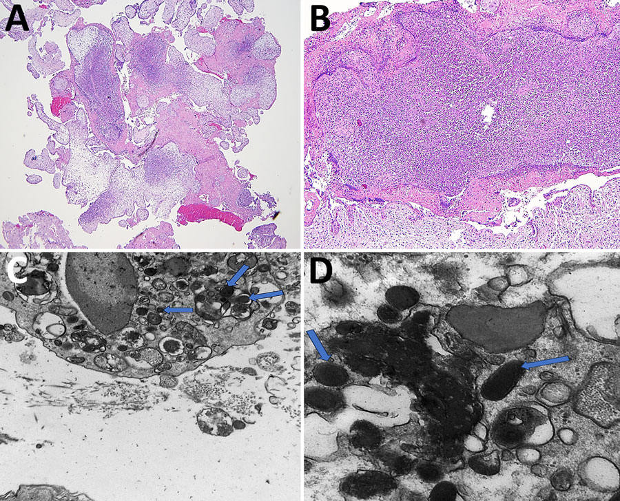
Figure 2. Imaging of samples from patient in study of infection by tickborne bacterium Candidatus Midichloria associated with first trimester pregnancy loss, Tennessee, USA. A, B) Formalin-fixed paraffin-embedded sections showing acute suppurative villitis and large intravillous abscesses. Original magnification ×40 for panel A and ×200 for panel B. C, D) Electron microscopy analysis was performed on tissue that was previously formalin-fixed but not paraffin-embedded. The formalin-fixed tissue was placed in a 2.5% glutaraldehyde solution before electron microscopy analysis. C) Intracellular bacterial forms in the cytosol (indicated by arrows) at ×20,000 magnification; D) cytoplasmic vacuoles (indicated by arrows) at ×60,000 magnification, measuring ≈0.25–0.34 μm × 0.40–0.53 μm.