Skip directly to site content Skip directly to page options Skip directly to A-Z link Skip directly to A-Z link Skip directly to A-Z link

Disclaimer: Early release articles are not considered as final versions. Any changes will be reflected in the online version in the month the article is officially released.

Volume 31, Number 3—March 2025
Dispatch

Simultaneous Detection of Sarcocystis hominis, S. heydorni, and S. sigmoideus in Human Intestinal Sarcocystosis, France, 2021–2024

Maxime Moniot, Patricia Combes, Damien Costa, Nicolas Argy, Marie-Fleur Durieux, Thomas Nicol, Céline Nourrisson, and Philippe PoirierComments to Author 
Author affiliation: CHU Clermont-Ferrand Service de Parasitologie-Mycologie, Clermont-Ferrand, France (M. Moniot); CHU Clermont-Ferrand Centre National de Référence des cryptosporidioses, microsporidies et autres protozooses digestives, Clermont-Ferrand (M. Moniot, P. Combes, D. Costa, C. Nourrisson, P. Poirier); Université Rouen Normandie, Rouen, France (D. Costa); CHU Rouen Centre National de Référence Cryptosporidioses, Microsporidies et autres protozooses digestives (Centre coordonnateur), Rouen, France (D. Costa); Hopital Bichat-Claude Bernard, Paris, France (N. Argy); MERIT UMR 216, IRD, Faculté de pharmacie de Paris, Université Paris Cité, Paris (N. Argy); Centre de biologie et de recherche en santé, Hôpital Universitaire Dupuytren, Limoges, France (M. Durieux); CHU Angers, Angers, France (T. Nicol); Microbes, Intestin, Inflammation et Susceptibilité de l’Hôte, UMR Inserm/Université Clermont Auvergne U1071, USC INRAE 1382, Clermont-Ferrand (C. Nourrisson, P. Poirier)

Main Article

Figure 1

Oocysts of Sarcocystis spp. from patients with human intestinal sarcocystosis, France, 2021–2024. A) Concentrated stool smear stained using the merthiolate-iodine-formaldehyde method. Sporulated oocysts are colorless and contain 2 elongated sporocysts. The oocyst wall is thin and often invisible in wet mount. B) Wet mount. Each sporocyst contains 4 banana-shaped sporozoites and a granular sporocyst residuum, which may be compact or dispersed. The 4 sporozoites are rarely seen in a single plane of focus. C) Fresh homogenized stool smear under fluorescent microscopy. Individual sporocysts are autofluorescents and will appear blue with an excitation filter of 330–365 nm. Scale bars indicate 10 µm.

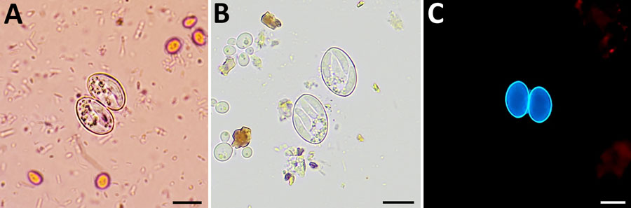
Figure 1. Oocysts of Sarcocystis spp. from patients with human intestinal sarcocystosis, France, 2021–2024. A) Concentrated stool smear stained using the merthiolate-iodine-formaldehyde method. Sporulated oocysts are colorless and contain 2 elongated sporocysts. The oocyst wall is thin and often invisible in wet mount. B) Wet mount. Each sporocyst contains 4 banana-shaped sporozoites and a granular sporocyst residuum, which may be compact or dispersed. The 4 sporozoites are rarely seen in a single plane of focus. C) Fresh homogenized stool smear under fluorescent microscopy. Individual sporocysts are autofluorescents and will appear blue with an excitation filter of 330–365 nm. Scale bars indicate 10 µm.

Main Article

Page created: February 04, 2025
Page updated: February 21, 2025
Page reviewed: February 21, 2025
The conclusions, findings, and opinions expressed by authors contributing to this journal do not necessarily reflect the official position of the U.S. Department of Health and Human Services, the Public Health Service, the Centers for Disease Control and Prevention, or the authors' affiliated institutions. Use of trade names is for identification only and does not imply endorsement by any of the groups named above.
file_external