Prospective Multicenter Surveillance of Non–H. pylori Helicobacter Infections during Medical Checkups, Japan
Kengo Tokunaga
1
, Emiko Rimbara
1 , Toshihisa Tsukadaira, Katsuhiro Mabe, Koji Yahara, Hidekazu Suzuki, Tadashi Shimoyama, Mitsushige Sugimoto, Tadayoshi Okimoto, Hidenori Matsui, Masato Suzuki, Keigo Shibayama, Hiroyoshi Ota, Kazunari Murakami, and Mototsugu Kato
Author affiliation: Kyorin University School of Medicine, Tokyo, Japan (K. Tokunaga); National Institute of Infectious Diseases, Tokyo (E. Rimbara, K. Yahara, H. Matsui, M. Suzuki); Kenwakai Hospital, Iida, Japan (T. Tsukadaira); Mabe Goryokaku Gastrointestinal Endoscopy Clinic, Hakodate, Japan (K. Mabe); Tokai University School of Medicine, Isehara, Japan (H. Suzuki); Aomori General Health Examination Center, Aomori, Japan (T. Shimoyama); Oita University, Oita, Japan (M. Sugimoto, K. Murakami); Oita Prefectural Hospital, Oita (T. Okimoto); Nagoya University Graduate School of Medicine, Nagoya, Japan (K. Shibayama); Shinshu University School of Medicine, Matsumoto, Japan (H. Ota); Public Interest Foundation Hokkaido Cancer Society, Sapporo, Japan (M. Kato)
Main Article
Figure 1
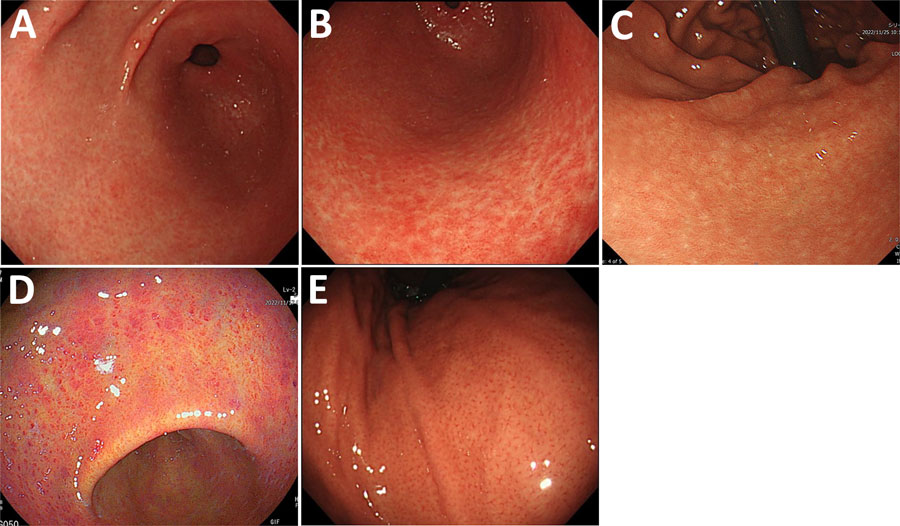
Figure 1. Representative endoscopic images of gastritis types observed in study of non–H. pylori Helicobacter infections, Japan, 2022. A) White marbled appearance. A white mesh pattern composed of shallow depressions that looks like white marble from the gastric antrum to the angle. White light imaging (WLI). B) Crack-like mucosa. A mesh pattern composed of faded and depressed lines with varied widths on coarse surfaces (WLI). C) Nodular gastritis. Compared with nodular gastritis caused by H. pylori infection, nodules that are shorter and appear as white spots are also considered nodular gastritis (WLI). D) Spotty redness, includes findings similar to those of H. pylori infection in the gastric body and the antrum (linked-color imaging). E) Regular arrangement of collecting venules. Microvessels with starfish-like appearance are observed as minute red points in the gastric lower body (WLI).
Main Article
Page created: March 31, 2025
Page updated: May 27, 2025
Page reviewed: May 27, 2025
The conclusions, findings, and opinions expressed by authors contributing to this journal do not necessarily reflect the official position of the U.S. Department of Health and Human Services, the Public Health Service, the Centers for Disease Control and Prevention, or the authors' affiliated institutions. Use of trade names is for identification only and does not imply endorsement by any of the groups named above.