Skip directly to site content Skip directly to page options Skip directly to A-Z link Skip directly to A-Z link Skip directly to A-Z link

Disclaimer: Early release articles are not considered as final versions. Any changes will be reflected in the online version in the month the article is officially released.

Volume 31, Number 8—August 2025

Research Letter

Emergence of Novel Fluoroquinolone Resistance Mutations in Mycoplasma bovis, China, 2008–2023

Shimei Lan, Shuang Liu, Wenjing Cui, Zhangcheng Li, Huafang Hao, Ahmed Adel Baz, Jinjia Liang, Xiangrui Jin, Xinmin Yan, Pengcheng Gao, Fuying Zheng, Shengli Chen1Comments to Author , and Yuefeng Chu1Comments to Author 
Author affiliation: State Key Laboratory for Animal Disease Control and Prevention, College of Veterinary Medicine, Lanzhou University, Lanzhou Veterinary Research Institute, Chinese Academy of Agricultural Sciences, Lanzhou, China (S. Lan, S. Liu, W. Cui, Z. Li, H. Hao, A.A. Baz, J. Liang, X. Jin, X. Yan, P. Gao, F. Zheng, S. Chen, Y. Chu); Gansu Province Research Center for Basic Disciplines of Pathogen Biology, Lanzhou (S. Lan, S. Liu, Z. Li, H. Hao, A.A. Baz, J. Liang, X. Jin, X. Yan, P. Gao, F. Zheng, S. Chen, Y. Chu); Key Laboratory of Veterinary Etiological Biology, Key Laboratory of Ruminant Disease Prevention and Control (West), Ministry of Agricultural and Rural Affairs, Lanzhou (S. Lan, S. Liu, Z. Li, H. Hao, A.A. Baz, J. Liang, X. Jin, X. Yan, P. Gao, F. Zheng, S. Chen, Y. Chu)

Main Article

Figure 2

Amino acid sequence alignments of quinolone resistance-determining regions of Mycoplasma bovis isolates from China, 2008–2023. Multiple alignments of conserved GyrA (A) and ParC (B) protein sequences for M. bovis ParC protein–ciprofloxacin complex are shown. Escherichia coli K12 and M. bovis PG45 strains were used as controls. Red arrows and black rectangular borders indicate amino acid mutation sites.

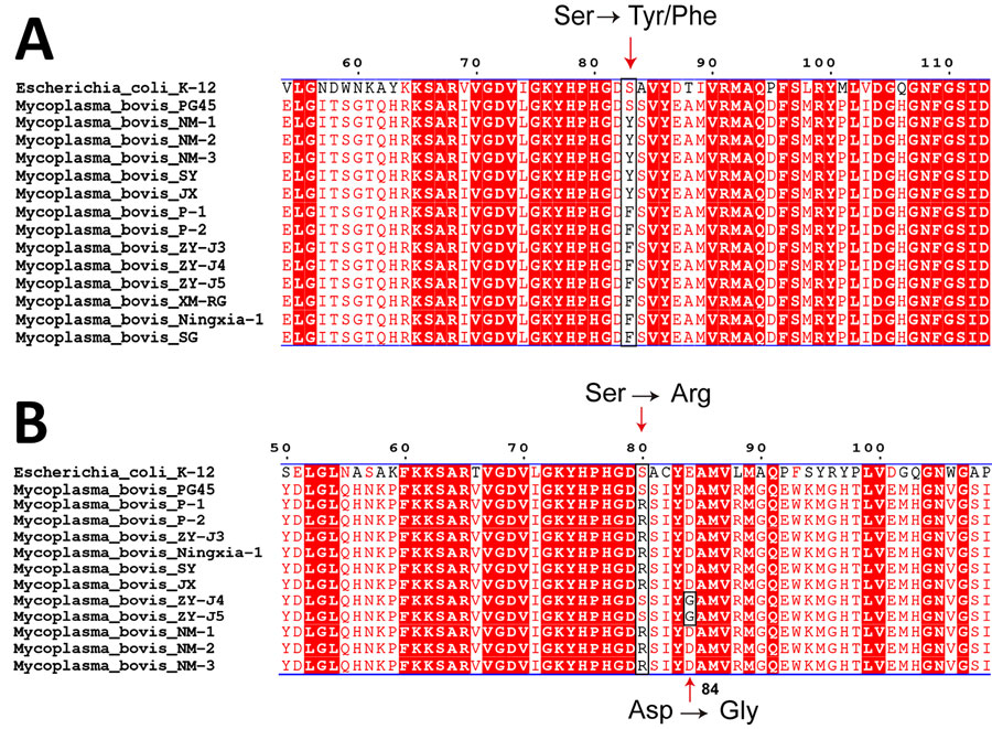
Figure 2. Amino acid sequence alignments of quinolone resistance-determining regions of Mycoplasma bovis isolates from China, 2008–2023. Multiple alignments of conserved GyrA (A) and ParC (B) protein sequences for M. bovis ParC protein–ciprofloxacin complex are shown. Escherichia coli K12 and M. bovis PG45 strains were used as controls. Red arrows and black rectangular borders indicate amino acid mutation sites.

Main Article

1These senior authors contributed equally to this article.

Page created: June 23, 2025
Page updated: July 16, 2025
Page reviewed: July 16, 2025
The conclusions, findings, and opinions expressed by authors contributing to this journal do not necessarily reflect the official position of the U.S. Department of Health and Human Services, the Public Health Service, the Centers for Disease Control and Prevention, or the authors' affiliated institutions. Use of trade names is for identification only and does not imply endorsement by any of the groups named above.
file_external