Skip directly to site content Skip directly to page options Skip directly to A-Z link Skip directly to A-Z link Skip directly to A-Z link

Disclaimer: Early release articles are not considered as final versions. Any changes will be reflected in the online version in the month the article is officially released.

Volume 32, Number 10—October 2026

Dispatch

Comparison of Baloxavir-Based Combinations and Monotherapies for Treating Influenza A(H5N1) Clade 2.3.4.4b Virus Infection in Mice

Seong Cheol Min, Ju Ryeong Lee, Beom Kyu Kim, Ji-Hyun Park, Dong Gyu Lee, Gi Chan Lee, Se Hee An, Santosh Chokkakula, Aman Jain, Young Ki Choi, Yun Hee Baek, and Min-Suk SongComments to Author 
Author affiliation: Chungbuk National University College of Medicine, Chungbuk, South Korea (S.C. Min, J.R. Lee, B.K. Kim, J.-H. Park, D.G. Lee, G.C. Lee, S.H. An, S. Chokkakula, A. Jain, Y.H. Baek, M.-S. Song); Korea Virus Research Institute Yuseong-gu, Daejeon, South Korea (Y.K. Choi)

Main Article

Figure 2

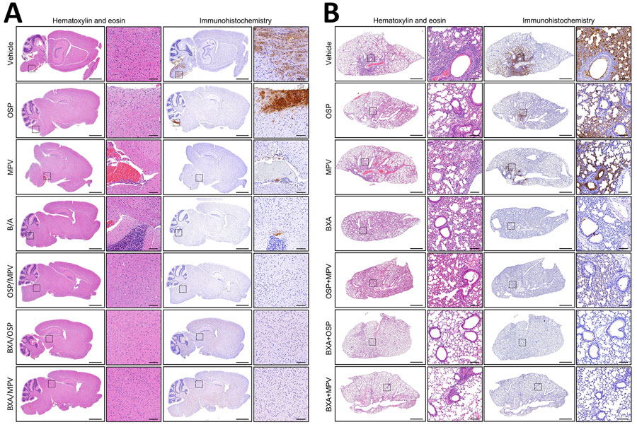
Lung and brain histopathology and influenza A nucleoprotein immunoreactivity at 6 dpi after monotherapy or combination therapy in a study of treating influenza A(H5N1) clade 2.3.4.4b virus infection in mice. A) Brain sections; B) lung sections. Sections from vehicle, OSP, MPV, BXA, OSP/MPV, BXA/OSP, and BXA/MPV groups. For each group, representative hematoxylin and eosin images (left) and immunohistochemistry for influenza A nucleoprotein (right; 3,3′-diaminobenzidine with hematoxylin counterstain) are shown as whole-section views with corresponding higher-magnification insets (boxed regions). Scale bars indicate 1.25 mm for whole-section views; and 100 μm for higher-magnification insets. BXA, baloxavir acid; MPV, molnupiravir; OSP, oseltamivir phosphate.

Figure 2. Lung and brain histopathology and influenza A nucleoprotein immunoreactivity at 6 dpi after monotherapy or combination therapy in a study of treating influenza A(H5N1) clade 2.3.4.4b virus infection in mice. A) Brain sections; B) lung sections. Sections from vehicle, OSP, MPV, BXA, OSP/MPV, BXA/OSP, and BXA/MPV groups. For each group, representative hematoxylin and eosin images (left) and immunohistochemistry for influenza A nucleoprotein (right; 3,3′-diaminobenzidine with hematoxylin counterstain) are shown as whole-section views with corresponding higher-magnification insets (boxed regions). Scale bars indicate 1.25 mm for whole-section views; and 100 μm for higher-magnification insets. BXA, baloxavir acid; MPV, molnupiravir; OSP, oseltamivir phosphate.

Main Article

Page created: September 05, 2026
Page updated: September 22, 2026
Page reviewed: September 22, 2026
The conclusions, findings, and opinions expressed by authors contributing to this journal do not necessarily reflect the official position of the U.S. Department of Health and Human Services, the Public Health Service, the Centers for Disease Control and Prevention, or the authors' affiliated institutions. Use of trade names is for identification only and does not imply endorsement by any of the groups named above.
file_external