Volume 32, Number 10—October 2026
Research
Persistent Bartonella quintana Infection in Macaques at Primate Research Institute, Japan

Cite This Article
Citation for Media
Abstract
Bartonella quintana, the causative agent of trench fever, is a louseborne bacterial pathogen. During 2016−2017, we investigated B. quintana infection in macaques housed at 2 primate research institutes in Japan. We isolated B. quintana from 3 wild-caught Japanese macaques, which had been introduced into 1 institute in 2009. The macaques had received periodic ivermectin treatment every 1–2 years to control blood-feeding arthropods. Whole-genome comparisons revealed that the 3 isolates exhibit higher homology to a Japanese macaque–derived strain than to rhesus macaque–derived or human-derived strains. In addition, in all 3 isolates, the bepA locus was absent; we classified the trwL locus as structural variant C, consistent with known Japanese macaque–derived strains. Our findings suggest that B. quintana bacteremia in the macaques might have persisted for 7–8 years in captivity, and that the bacterium was not accidentally transmitted to them from humans by reverse zoonosis.
The genus Bartonella belongs to the class Alphaproteobacteria, order Rhizobiales, and family Bartonellaceae and currently comprises 49 species and 3 subspecies (1). Bartonella bacteria are transmitted among their natural hosts by blood-feeding arthropods, including fleas, lice, keds, and sand flies, which excrete the bacteria in their feces (2). Mammals, such as bats, cats, deer, rabbits, and rodents serve as natural reservoirs for Bartonella species (3–6). Vascular endothelial cells are the initial cellular targets during Bartonella infection, after which the bacteria are released into the host bloodstream and adhere to erythrocytes. Adherence of Bartonella bacteria to erythrocytes is mediated through the Trw type IV secretion system, a key determinant of host specificity among Bartonella species (7). Once established within erythrocytes, Bartonella bacteria are thought to persist as long-term intraerythrocytic parasites.
B. quintana is the etiologic agent of trench fever, a human infectious disease transmitted by the human body louse. Trench fever was first documented among Allied and German troops during World War I and sporadically reemerged during World War II (8). In immunocompetent persons, the disease typically manifests as relapsing fever for several days accompanied by bacteremia, severe headache, dizziness, and persistent pain in the tibias. More severe manifestations, including bacillary angiomatosis and infective endocarditis, have been reported in immunocompromised patients, particularly those with human immunodeficiency virus infection (8). Epidemiologic studies have demonstrated that B. quintana infection occurs predominantly among homeless populations in urban settings in the Americas, Europe, and Asia (9–13). Consequently, B. quintana is regarded as a reemerging pathogen found in modern societies.
Although those symptoms are observed in trench fever patients, B. quintana infection typically manifests as an asymptomatic condition in which the bacteria harbor within erythrocytes (14). A previous study conducted in Marseille, France, showed that 10/71 homeless persons tested positive for B. quintana bacteremia; 6 of those were afebrile (10). The remaining 4 patients exhibited prolonged bacteremia, persisting for 4–6 weeks. Asymptomatic carriers with B. quintana have been identified in other homeless populations in Marseille (11); 1 person exhibited chronic B. quintana bacteremia for 78 weeks, and 2 others remained bacteremic for 17 and 53 weeks. In the 1930s and 1940s, the first reports of chronic B. quintana bacteremia emerged among employees of laboratories producing Weigl’s type vaccines against typhus in Poland. As reported in the literature, B. quintana has been isolated from those employees for as long as 8 years (15). Subsequently, chronic bacteremia was demonstrated in experimental studies in the 1960s in which human volunteers were inoculated with the bacterium (16). From those findings, the duration of B. quintana bacteremia in the carriers is estimated to range from several weeks to ≈8 years.
Macaques inhabiting Asia have been identified as additional hosts for B. quintana. The first reported case of B. quintana infection in macaques was in a captive-bred cynomolgus macaque (Macaca fascicularis) imported into the United States from Vietnam (17). Subsequently, the bacterium was isolated from cynomolgus macaques and rhesus macaques (M. mulatta) born and reared at primate research centers in China (18,19) as well as from wild-caught Japanese macaques (M. fuscata) in Japan (20). In a previous study (20), no clinical signs were observed in the Japanese macaques positive for B. quintana infection, as seen in human patients with chronic B. quintana bacteremia; however, the duration of bacteremia in the macaques remains unknown.
A comparative genomic analysis of complete B. quintana genomes has revealed notable genomic differences among B. quintana strains from human, rhesus macaque, and Japanese macaque (21). Among those strains, the Japanese macaque–derived strains exhibit unique genomic features not observed in the others. Of note, strains MF1-1, MF3-1, MF10-1, MF11-1, MF19-1, and MF34-1 lack bepA, a gene known to confer anti-apoptotic effects in vascular endothelial cells. In addition, those strains harbor novel genomic structures within the trwL locus, which is implicated in erythrocyte adhesion. The human-derived strain Toulouse and the rhesus macaque–derived strain RM-11 share a conserved trwL structure, comprising trwL1 to trwL8, in common. On the other hand, 3 distinct structural variants (A–C) have been identified among Japanese macaque strains; variant A consists of the gene order trwL1–trwL5–trwL3–trwL7–trwL8, variant B of trwL1–trwLx–trwL5–trwL7–trwL8, and variant C of trwL1–trwLx–trwL5–trwL3–trwL7–trwL8. The presence of trwLx, newly identified between trwL1 and trwL5 in variants B and C, highlights substantial genomic diversity within the trwL locus even among Japanese macaque strains (21).
Although the prevalence of B. quintana infection and the genomic properties of Bartonella in wild-caught Japanese macaques have been documented, its occurrence in experimental macaques controlled by human management in Japan remains unknown. In this study, we investigated B. quintana infection in macaque populations maintained at 2 primate research institutes in Japan. Furthermore, we performed whole-genome sequencing of B. quintana strains from the infected macaques to genetically characterize the bepA and trwL loci.
Blood Sampling
During 2016−2017, blood samples were aseptically collected from 92 wild-caught Japanese macaques and from 336 captive-bred macaques including 235 Japanese macaques and 101 rhesus macaques at the Primate Research Institute (PRI), Kyoto University (Inuyama, Japan). In 2017, additional blood samples were aseptically collected from 90 captive-bred cynomolgus macaques at the Tsukuba Primate Research Center (TPRC), National Institutes of Biomedical Innovation, Health, and Nutrition (Tsukuba, Japan) (Table 1). All blood samples were immediately stored at −70°C until further analysis.
The wild-caught Japanese macaques originated from several geographic locations and were transferred to PRI as follows: 8 macaques from Aichi Prefecture, in August 2003; 15 from Shizuoka Prefecture, in June 2008; 22 from Wakayama Prefecture in February 2009; and 47 from Osaka Prefecture during March 2008–June 2009. Upon arrival at the facility, all wild-caught macaques underwent deworming treatments with ivermectin. The ivermectin solution Ivermec Inj (Fujita Pharmaceutical Co., http://www3.fujita-pharm.co.jp) was administered subcutaneously at a dose of 0.1 mL/kg. In addition, both wild-caught and captive-bred macaques received periodic ivermectin treatments during routine physical examinations conducted at intervals of 1–2 years. At PRI, macaques were kept outdoors in social groups according to their geographic origin. In contrast, macaques at the TPRC were housed indoors in individual cages and received medicated baths with the organophosphorus ectoparasiticide Neguvon (Bayer, https://www.bayer.com) at 2-year intervals.
All animal procedures were conducted in accordance with applicable animal welfare regulations. Blood sampling at the PRI was performed under approved animal experimentation protocols (approval nos. 2016-158 and 2017-089). Blood samples from the TPRC were provided through the Nonhuman Primate Reagent and Resource Program under an institutional agreement for the distribution of research materials.
Detection of Bartonella DNA from Macaque Blood Samples
To screen for B. quintana infection in the macaque populations, we performed nested PCR targeting the RNA polymerase β-subunit gene (rpoB) of the genus Bartonella. We designed the first PCR primers in this study, 1913f and 2357r, based on the rpoB sequences from B. quintana strains MF1-1 and RM-11. We designed the second PCR primers, 600f-m and 800r-m, with some modification based on original primers (22) (Table 2). The positive control was genomic DNA extracted from B. quintana strain MF1-1, whereas the negative control was genomic DNA extracted from a blood of a cynomolgus macaque classified as SPF grade. Conditions for the nested PCR were as follows: 2 minutes at 94°C followed by 35 cycles of 30 seconds at 94°C, 30 seconds at 54°C, and 1 minutes at 72°C, and a final extension of 2 minutes at 72°C. We purified the second PCR amplicon using a Wizard SV Gel and PCR Clean-Up System (Promega Corporation, https://www.promega.com), followed by direct DNA sequencing with the Genetic Analyzer models 3730xl (Thermo Fisher Scientific, https://www.thermofisher.com). We compared the obtained rpoB sequences with those in the International Nucleotide Sequence Database using the BLAST (https://blast.ncbi.nlm.nih.gov/Blast.cgi).
Isolation and Identification of Bartonella Bacteria from Macaque Bloods
We isolated Bartonella bacteria as described previously (20). In brief, we spread a 100 µL of freeze-thawed blood samples onto 5% rabbit blood chocolate agar plates and incubated them at 35°C under 5% CO2 for up to 4 weeks. We tentatively identified colonies as Bartonella species on the basis of characteristic morphology (small, round, gray to cream-yellow colonies) and the requirement for a long culture period (>1 week). We calculated colony-forming units (CFUs) per milliliter of blood. We recovered 5 colonies from each culture-positive sample and subcultured them on fresh chocolate agar plates under the same conditions as the primary culture.
We extracted genomic DNA from each isolate using InstaGene Matrix (Bio-Rad Laboratories, https://www.bio-rad.com). We performed species identification by Bartonella-specific PCR targeting the rpoB (23), citrate synthase (gltA) (24), and 16S–23S rDNA intergenic transcribed spacer (ITS) regions (25). We used genomic DNA from B. alsatica strain IBS382T as the positive control and nuclease-free distilled water as the negative control. We purified and sequenced PCR amplicons and compared them with the corresponding sequences derived from B. quintana strain MF1-1 (RefSeq [https://www.ncbi.nlm.nih.gov/refseq] accession no. NZ_AP019773).
Whole-Genome Sequencing Analysis
We extracted genomic DNAs from representative strains using DNeasy Blood and Tissue Kit (QIAGEN, https://www.qiagen.com). We prepared sequencing DNA libraries from the extracted genomic DNAs by using Illumina DNA Prep and IDT for Illumina DNA-RNA UD Indexes Set A, Tagmentation (Illumina, https://www.illumina.com). We sequenced the DNA libraries on a MiSeq platform using the MiSeq Reagent Nanokit version 2 (500 cycle) (Illumina). We quality-checked raw reads and assembled them using a de novo assembler program, the strategic K-mer extension for scrupulous assemblies (26), to generate contigs. We assessed de novo assembly qualities using Benchmarking Universal Single-Copy Orthologs (BUSCO) version 5.4.3 (27).
We assessed nucleotide sequence identities at whole-genome levels by pairwise average nucleotide identity (ANI) (28) and digital DNA-DNA hybridization (dDDH) (29). We compared the assemblies of representative strains with strains MF1-1, RM-11, and Toulouse.
We confirmed the presence or absence of the bepA locus comprising bepA1 and bepA2 by bepA-specific PCR and subsequently validated by whole-genome sequencing data. We identified structural variants within the trwL locus by comparison with previously reported data (21).
Data Availability
We deposited raw reads of all the sequenced B. quintana strains in this study in the Sequence Read Archive of the DNA Data Bank of Japan. All are available under BioProject accession no. PRJDB40655.
Prevalence of B. quintana in Macaque Populations at PRI and TPRC
We confirmed Bartonella infection by the rpoB-targeting PCR in 3 (3.3%) (sample IDs TB1, MN51, and MN57) of the 92 wild-caught Japanese macaques housed at PRI. All PCR amplicons showed an identical sequence to the rpoB of strain MF1-1. In contrast, we detected no Bartonella DNA in the captive-bred macaques at either the PRI or the TPRC.
We isolated Bartonella bacteria from the 3 PCR-positive Japanese macaques but not from any of the captive-bred macaques we examined. Bacteremia levels were 2.9 × 10³ CFU/mL for TB1, 4.0 × 10² CFU/mL for MN51, and 3.0 × 10³ CFU/mL for MN57. We recovered a total of 15 Bartonella isolates, 5 isolates per Bartonella-positive macaque. All the isolates showed 100% sequence identity for the gltA and rpoB and 99.9%–100% sequence identity for the ITS, compared with those of strain MF1-1.
According to management records at PRI, macaque TB1 was captured in Wakayama Prefecture and transferred to the PRI in February 2009. Similarly, macaques MN51 and MN57 were captured in Osaka Prefecture and transferred to PRI in June 2009. Blood samples for this study were collected in November 2016 from TB1 and in November 2017 from MN51 and MN57.
De Novo Assemblies Using Whole-Genome Sequences
We randomly selected 1 representative strain from each culture-positive macaque, yielding strains TB1-1, MN51-1, and MN57-1 for whole-genome sequencing analysis. The number of sequencing reads generated by the MiSeq platform ranged from 303,794 for strain MN51-1 to 1,065,204 for strain MN57-1 (Table 3). De novo assembly produced 43 contigs for strain TB1-1, 52 contigs for strain MN51-1, and 47 contigs for strain MN57-1; average genome coverage exceeded 46×. We estimated genome sizes of the 3 strains on the basis of the assembled contigs as ≈1.6 Mbp with guanine-cytosine contents of 38.8% for all strains. BUSCO scores of the 3 assembled contigs were 97.8–97.9.
We compared whole-genome sequences by ANI and dDDH. We compared the 3 representative strains with strain MF1-1 from a Japanese macaque, strain RM-11 from a rhesus macaque, and strain Toulouse from a human (Table 4). The ANI values obtained from the comparison of the representative 3 strains with strain MF1-1 were 99.8%, higher than those against strain RM-11 (99.4%) and strain Toulouse (98.3%). Similarly, the dDDH values obtained from comparing the representative strains with strain MF1-1 were 98.9%–99.0%, which exceeded those against strain RM-11 (95.3%–95.4%) and strain Toulouse (85.2%–85.3%).
Genomic Characterization of the bepA and trwL Loci
Figure 1
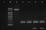
Figure 1. Electrophoretic analysis of bepA-specific PCR amplicons in study of persistent Bartonella quintanainfection in macaques at primate research institute, Japan. Lane M, 0.1–2 kb DNA size marker;...
Figure 2
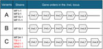
Figure 2. Structural variants of the trwL locus among Bartonella quintanastrains from Japanese macaques in study at primate research institute, Japan. We reviewed 3 structural variants (A–C) of...
The bepA locus (1,612bp) was absent from all 3 representative strains (Figure 1), which we confirmed by the whole-genome sequencing data. Gene synteny analysis revealed that the trwL locus in the representative strains corresponded to structural variant C, characterized by the gene order of trwL1, trwLx, trwL5, trwL3, trwL7, and trwL8 (Figure 2).
In this study, we isolated B. quintana in a research colony of 3 Japanese macaques housed at the PRI, all of which originated from wild-caught populations. The bacteremia levels (400–3,000 CFU/mL) were comparable to those previously reported in Japanese macaques (20). In contrast, no Bartonella infection was detected in any of the captive-bred Japanese, cynomolgus, or rhesus macaques housed at either of the 2 primate facilities. Although both wild-caught and captive-bred macaques at the 2 facilities received periodic antiparasitic treatments, the B. quintana–positive Japanese macaques might have been infested with some kind of blood-feeding arthropods, such as monkey lice (Pedicinus sp.) (30), before they were captured in their habitats. We found monkey lice on wild-caught Japanese macaques in Kanagawa Prefecture, Japan, during 2018–2022 and successfully detected B. quintana DNA from the lice (data not shown). Because the B. quintana–positive Japanese macaques were introduced into the PRI in 2009 and tested positive for B. quintana infection in 2016–2017, we presumed that they maintained persistent B. quintana infection for up to 8 years. Similarly, in 1949, B. quintana bacteremia was documented as persisting for up to 8 years in trench fever patients (15). On the basis of all those findings, we concluded that B. quintana can establish long-term persistent infections in both humans and nonhuman primates. Such long-term, asymptomatic B. quintana bacteremia demonstrates that infected individuals can act as chronic carriers of the pathogen without manifesting distinct clinical symptoms. However, we noted that chronic bacteremic states in humans might eventually progress to severe complications, such as endocarditis (31).
We performed subsequent whole-genome sequencing to genetically characterize the 3 representative strains TB1-1, MN51-1, and MN57-1 from the B. quintana–positive Japanese macaques. We evaluated genome assembly quality using BUSCO analysis, which yielded completeness scores >95% for all strains (27), supporting the reliability of the de novo assemblies. In addition, the estimated genome sizes and guanine-cytosine contents were highly consistent with those reported for other known B. quintana strains (21,32), indicating that the assembled genomes were suitable for downstream comparative analyses. Comparisons at the whole-genome level using ANI and dDDH revealed that the 3 strains exhibit higher homology to strain MF1-1 from a Japanese macaque (21) than to strain RM-11 from a rhesus macaque and strain Toulouse from a human. Of note, in all 3 strains, the bepA locus was absent, and the trwL locus was classified as structural variant C; we had demonstrated previously that both loci seem to be unique to Japanese macaque–derived strains (21). Those findings suggest that the B. quintana–positive Japanese macaques did not accidentally acquire the bacterium from humans within this facility, ruling out the likelihood that reverse zoonosis caused the infections. Future surveillance of B. quintana infection in other nonhuman primates, such as apes and New World monkeys, will increase understanding of the host range of the bacterium in nature.
Studying infection state in the same animals over time would be helpful to further confirm long-term persistent infection. However, the PRI ethics guidelines restrict blood sampling from the same macaques to once every 2 years. Furthermore, the macaque TB1 died in June 2018 from an accident within the PRI enclosure, which prevented the collection of consecutive blood samples. Although we attempted to collect additional blood samples from macaques MN51 and MN57 since November 2019, experiments at PRI were suspended for several years during the COVID-19 pandemic, making subsequent medical research applications exceedingly challenging. To address those issues, we would need to conduct infection experiments for Macaca spp. monkeys with Japanese macaque–derived strains in another primate research facility.
In conclusion, we identified B. quintana bacteremia in 3 wild-caught Japanese macaques housed at PRI and noted evidence suggesting persistent infection for up to 8 years after capture. Given that the trw operon, particularly the trwL and trwJ loci, plays a crucial role in erythrocyte adhesion (7), further investigation is warranted to determine whether structural variations in trwL influence the ability of macaque-derived B. quintana strains to infect human erythrocytes. Such studies would be particularly relevant from One Health perspective. Our findings highlight the capacity of B. quintana bacteria to establish long-term infection in Japanese macaques and support previous documentation of prolonged bacteremia in humans.
Mr. Nomura is a PhD candidate graduate student in the Department of Veterinary Medicine, College of Bioresource Sciences, Nihon University, Japan. His primary research interests are the epidemiology, elucidation of the virulence mechanisms, and prevention of bacterial zoonoses.
Acknowledgment
This work was supported in part by the Japan Society for the Promotion of Science Grant-in-Aid for Young Scientists (B) (grant no. 17K15373) and the Cooperative Research Program of Primate Research Institute, Kyoto University (2016 to 2018).
References
- List of prokaryotic names with standing in nomenclature. Genus Bartonella [cited 2026 Jan 11]. https://lpsn.dsmz.de/genus/Bartonella
- Tsai YL, Chang CC, Chuang ST, Chomel BB. Bartonella species and their ectoparasites: selective host adaptation or strain selection between the vector and the mammalian host? Comp Immunol Microbiol Infect Dis. 2011;34:299–314. DOIPubMedGoogle Scholar
- Qiu Y, Kajihara M, Nakao R, Mulenga E, Harima H, Hang’ombe BM, et al. Isolation of Candidatus Bartonella rousetti and other bat-associated Bartonellae from bats and their flies in Zambia. Pathogens. 2020;9:469. DOIPubMedGoogle Scholar
- Ciceroni L, Fabbi M, Ciarrocchi S, Pinto A, Ciervo A, Kasten RW, et al. Characterization of the first Bartonella henselae strain isolated from a cat in Italy. Comp Immunol Microbiol Infect Dis. 2002;25:217–28. DOIPubMedGoogle Scholar
- Sato S, Kabeya H, Yamazaki M, Takeno S, Suzuki K, Kobayashi S, et al. Prevalence and genetic diversity of Bartonella species in sika deer (Cervus nippon) in Japan. Comp Immunol Microbiol Infect Dis. 2012;35:575–81. DOIPubMedGoogle Scholar
- Gonçalves-Oliveira J, Gutierrez R, Schlesener CL, Jaffe DA, Aguilar-Setién A, Boulouis HJ, et al. Genomic characterization of three novel Bartonella strains in a rodent and two bat species from Mexico. Microorganisms. 2023;11:340. DOIPubMedGoogle Scholar
- Deng H, Pang Q, Zhao B, Vayssier-Taussat M. Molecular mechanisms of Bartonella and mammalian erythrocyte interactions: a review. Front Cell Infect Microbiol. 2018;8:431. DOIPubMedGoogle Scholar
- Maurin M, Raoult D. Bartonella (Rochalimaea) quintana infections. Clin Microbiol Rev. 1996;9:273–92. DOIPubMedGoogle Scholar
- Spach DH, Kanter AS, Dougherty MJ, Larson AM, Coyle MB, Brenner DJ, et al. Bartonella (Rochalimaea) quintana bacteremia in inner-city patients with chronic alcoholism. N Engl J Med. 1995;332:424–8. DOIPubMedGoogle Scholar
- Brouqui P, Lascola B, Roux V, Raoult D. Chronic Bartonella quintana bacteremia in homeless patients. N Engl J Med. 1999;340:184–9. DOIPubMedGoogle Scholar
- Foucault C, Barrau K, Brouqui P, Raoult D. Bartonella quintana bacteremia among homeless people. Clin Infect Dis. 2002;35:684–9. DOIPubMedGoogle Scholar
- Capo C, Amirayan-Chevillard N, Brouqui P, Raoult D, Mege JL. Bartonella quintana bacteremia and overproduction of interleukin-10: model of bacterial persistence in homeless people. J Infect Dis. 2003;187:837–44. DOIPubMedGoogle Scholar
- Sasaki T, Adachi T, Itoh K, Matsuoka M, Yamagishi T, Hirao M, et al. Detection of Bartonella quintana infection among the homeless population in Tokyo, Japan, from 2013–2015. Jpn J Infect Dis. 2021;74:411–5. DOIPubMedGoogle Scholar
- Rolain JM, Foucault C, Guieu R, La Scola B, Brouqui P, Raoult D. Bartonella quintana in human erythrocytes. Lancet. 2002;360:226–8. DOIPubMedGoogle Scholar
- Kostrzewski J. The epidemiology of trench fever [in Polish]. Bull Int Acad Pol Sci Let Cl Med. 1949;7:233–63.PubMedGoogle Scholar
- Vinson JW, Varela G, Molina-Pasquel C. Trench fever. Am J Trop Med Hyg. 1969;18:713–22. DOIPubMedGoogle Scholar
- O’Rourke LG, Pitulle C, Hegarty BC, Kraycirik S, Killary KA, Grosenstein P, et al. Bartonella quintana in cynomolgus monkey (Macaca fascicularis). Emerg Infect Dis. 2005;11:1931–4. DOIPubMedGoogle Scholar
- Huang R, Liu Q, Li G, Li D, Song X, Birtles RJ, et al. Bartonella quintana infections in captive monkeys, China. Emerg Infect Dis. 2011;17:1707–9. DOIPubMedGoogle Scholar
- Li H, Bai JY, Wang LY, Zeng L, Shi YS, Qiu ZL, et al. Genetic diversity of Bartonella quintana in macaques suggests zoonotic origin of trench fever. Mol Ecol. 2013;22:2118–27. DOIPubMedGoogle Scholar
- Sato S, Kabeya H, Yoshino A, Sekine W, Suzuki K, Tamate HB, et al. Japanese macaques (Macaca fuscata) as natural reservoir of Bartonella quintana. Emerg Infect Dis. 2015;21:2168–70. DOIPubMedGoogle Scholar
- Sato S, Nishioka E, Kabeya H, Maruyama S. Genomic properties of a Bartonella quintana strain from Japanese macaque (Macaca fuscata) revealed by genome comparison with human and rhesus macaque strains. Sci Rep. 2024;14:10941. DOIPubMedGoogle Scholar
- Gutiérrez R, Morick D, Gross I, Winkler R, Abdeen Z, Harrus S. Bartonellae in domestic and stray cats from Israel: comparison of bacterial cultures and high-resolution melt real-time PCR as diagnostic methods. Vector Borne Zoonotic Dis. 2013;13:857–64. DOIPubMedGoogle Scholar
- Kabeya H, Inoue K, Izumi Y, Morita T, Imai S, Maruyama S. Bartonella species in wild rodents and fleas from them in Japan. J Vet Med Sci. 2011;73:1561–7. DOIPubMedGoogle Scholar
- Norman AF, Regnery R, Jameson P, Greene C, Krause DC. Differentiation of Bartonella-like isolates at the species level by PCR-restriction fragment length polymorphism in the citrate synthase gene. J Clin Microbiol. 1995;33:1797–803. DOIPubMedGoogle Scholar
- Roux V, Raoult D. The 16S-23S rRNA intergenic spacer region of Bartonella (Rochalimaea) species is longer than usually described in other bacteria. Gene. 1995;156:107–11. DOIPubMedGoogle Scholar
- Souvorov A, Agarwala R, Lipman DJ. SKESA: strategic k-mer extension for scrupulous assemblies. Genome Biol. 2018;19:153. DOIPubMedGoogle Scholar
- Simão FA, Waterhouse RM, Ioannidis P, Kriventseva EV, Zdobnov EM. M Simão FA. BUSCO: assessing genome assembly and annotation completeness with single-copy orthologs. Bioinformatics. 2015;31:3210–2. DOIPubMedGoogle Scholar
- Richter M, Rosselló-Móra R, Oliver Glöckner F, Peplies J. JSpeciesWS: a web server for prokaryotic species circumscription based on pairwise genome comparison. Bioinformatics. 2016;32:929–31. DOIPubMedGoogle Scholar
- Meier-Kolthoff JP, Carbasse JS, Peinado-Olarte RL, Göker M. TYGS and LPSN: a database tandem for fast and reliable genome-based classification and nomenclature of prokaryotes. Nucleic Acids Res. 2022;50(D1):D801–7. DOIPubMedGoogle Scholar
- Li H, Liu W, Zhang GZ, Sun ZZ, Bai JY, Jiang BG, et al. Transmission and maintenance cycle of Bartonella quintana among rhesus macaques, China. Emerg Infect Dis. 2013;19:297–300. DOIPubMedGoogle Scholar
- Okaro U, Addisu A, Casanas B, Anderson B. Bartonella species, an emerging cause of blood-culture-negative endocarditis. Clin Microbiol Rev. 2017;30:709–46. DOIPubMedGoogle Scholar
- Alsmark CM, Frank AC, Karlberg EO, Legault BA, Ardell DH, Canbäck B, et al. The louse-borne human pathogen Bartonella quintana is a genomic derivative of the zoonotic agent Bartonella henselae. Proc Natl Acad Sci U S A. 2004;101:9716–21. DOIPubMedGoogle Scholar
Figures
Tables
Cite This ArticleOriginal Publication Date: September 17, 2026
Table of Contents – Volume 32, Number 10—October 2026
| EID Search Options |
|---|
 Advanced Article Search – Search articles by author and/or keyword.
|
 Articles by Country Search – Search articles by the topic country.
|
 Article Type Search – Search articles by article type and issue.
|
Please use the form below to submit correspondence to the authors or contact them at the following address:
Shingo Sato, Nihon University—Veterinary Medicine,1866 Kameino, Fujisawa Kanagawa 252-0880, Japan
Top