Skip directly to site content Skip directly to page options Skip directly to A-Z link Skip directly to A-Z link Skip directly to A-Z link

Volume 32, Number 2—February 2026

Research

Pulmonary Complications in Fatal Yellow Fever, Brazil, 2017–2019

Amaro N. Duarte-NetoComments to Author , Katia C. Dantas, Suzette C. Ferreira, Fernando R. Giugni, Marielton P. Cunha, Shahab Z. Pour, Felipe L. Ledesma, Yeh-Li Ho, Ana C.S. Nastri, Cinthya S.C. Borges, Fernanda A. Rodrigues, Ceila M.S. Malaque, Jaques Sztajnbok, Thais Mauad, Luiz F.F. Silva, Paulo H.N. Saldiva, and Marisa Dolhnikoff
Author affiliation: Universidade de São Paulo, São Paulo, São Paulo, Brazil (A.N. Duarte-Neto, K.C. Dantas, F.R. Giugni, M.P. Cunha, S.Z. Pour, F.L. Ledesma, Y.-L. Ho, A.C.S. Nastri, F.A. Rodrigues, T. Mauad, L.F.F. Silva, P.H.N. Saldiva, M. Dolhnikoff); Fundação Pró-Sangue Hemocentro de São Paulo, São Paulo (S.C. Ferreira); Instituto Adolfo Lutz, São Paulo (C.S.C. Borges); Instituto de Infectologia Emílio Ribas, São Paulo (C.M.S. Malaque, J. Sztajnbok).

Main Article

Figure 3

Pulmonary vascular damage in fatal yellow fever cases, 2017–2019 epidemic, São Paulo, Brazil. A) Medium-sized artery with fibrinoid necrosis of the endothelial layer, marginated leukocytes, wall edema, septal congestion, alveolar hemorrhage. Hematoxylin and eosin (HE) stain; scale bar 50 µm. Inset shows group of hemosiderin-laden alveolar macrophages stained for iron. Perls stain; original magnification ´200. B) Pulmonary artery showing angioinvasion by Aspergillus spp. forming fibrinous thrombus on the endothelial vascular layer (black arrow). HE stain; scale bar = 50 µm. C) A small fibrin clot and the artery fibrinoid necrosis and wall edema. Periodic acid–Schiff stain; scale bar = 50 µm. D) Positive detection of VIII coagulation factor in the entire wall of pulmonary arteries. Peroxidase stain; scale bar = 20 µm. E) The VCAM is detected in the endothelial and muscular pulmonary artery layers. Peroxidase stain; scale bar = 20 µm. F) Septal capillaries showing mitochondrial dilation with loss of cristae. Ultrathin section; scale bar = 1 µm. G) Bacilli (arrows) within septal pulmonary vessel. Ultrathin section; scale bar = 500 nm. H) Histologic leukostasis in a septal pulmonary artery, showing immature myeloid cells, lymphocytes, and neutrophils. HE stain; scale bar = 50 µm. I) Intravascular cells expressing yellow fever virus antigens in their cytoplasm. Alkaline phosphatase stain; scale bar = 50 µm. J) The yellow fever virus antigen is detected in the cytoplasm of septal endothelial cells (inset; original magnification ´400) and in interstitial and alveolar inflammatory cells. Alkaline phosphatase stain; scale bar = 20 µm.

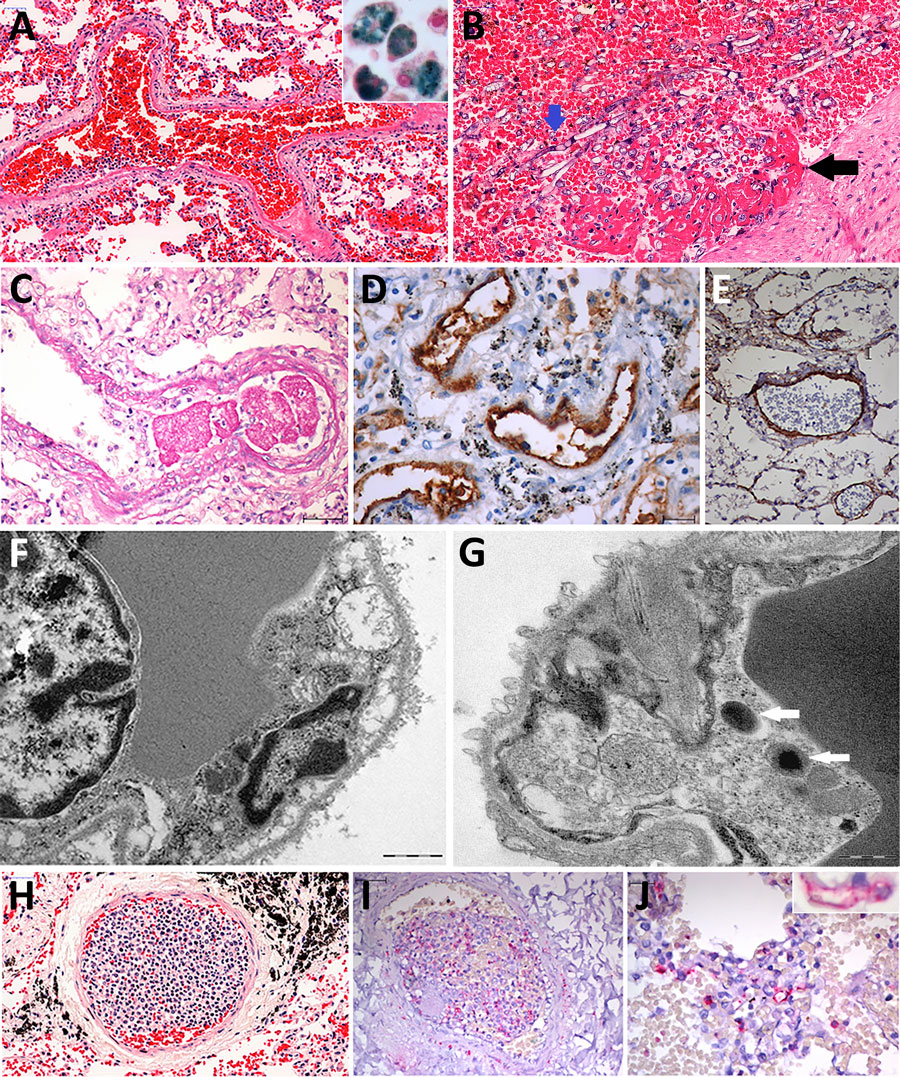
Figure 3. Pulmonary vascular damage in fatal yellow fever cases, 2017–2019 epidemic, São Paulo, Brazil. A) Medium-sized artery with fibrinoid necrosis of the endothelial layer, marginated leukocytes, wall edema, septal congestion, alveolar hemorrhage. Hematoxylin and eosin (HE) stain; scale bar 50 µm. Inset shows group of hemosiderin-laden alveolar macrophages stained for iron. Perls stain; original magnification ´200. B) Pulmonary artery showing angioinvasion by Aspergillus spp. forming fibrinous thrombus on the endothelial vascular layer (black arrow). HE stain; scale bar = 50 µm. C) A small fibrin clot and the artery fibrinoid necrosis and wall edema. Periodic acid–Schiff stain; scale bar = 50 µm. D) Positive detection of VIII coagulation factor in the entire wall of pulmonary arteries. Peroxidase stain; scale bar = 20 µm. E) The VCAM is detected in the endothelial and muscular pulmonary artery layers. Peroxidase stain; scale bar = 20 µm. F) Septal capillaries showing mitochondrial dilation with loss of cristae. Ultrathin section; scale bar = 1 µm. G) Bacilli (arrows) within septal pulmonary vessel. Ultrathin section; scale bar = 500 nm. H) Histologic leukostasis in a septal pulmonary artery, showing immature myeloid cells, lymphocytes, and neutrophils. HE stain; scale bar = 50 µm. I) Intravascular cells expressing yellow fever virus antigens in their cytoplasm. Alkaline phosphatase stain; scale bar = 50 µm. J) The yellow fever virus antigen is detected in the cytoplasm of septal endothelial cells (inset; original magnification ´400) and in interstitial and alveolar inflammatory cells. Alkaline phosphatase stain; scale bar = 20 µm.

Main Article

Page created: January 20, 2026
Page updated: February 19, 2026
Page reviewed: February 19, 2026
The conclusions, findings, and opinions expressed by authors contributing to this journal do not necessarily reflect the official position of the U.S. Department of Health and Human Services, the Public Health Service, the Centers for Disease Control and Prevention, or the authors' affiliated institutions. Use of trade names is for identification only and does not imply endorsement by any of the groups named above.
file_external