Volume 32, Number 2—February 2026
Research
Pulmonary Complications in Fatal Yellow Fever, Brazil, 2017–2019
Amaro N. Duarte-Neto , Katia C. Dantas, Suzette C. Ferreira, Fernando R. Giugni, Marielton P. Cunha, Shahab Z. Pour, Felipe L. Ledesma, Yeh-Li Ho, Ana C.S. Nastri, Cinthya S.C. Borges, Fernanda A. Rodrigues, Ceila M.S. Malaque, Jaques Sztajnbok, Thais Mauad, Luiz F.F. Silva, Paulo H.N. Saldiva, and Marisa Dolhnikoff
Figure 1
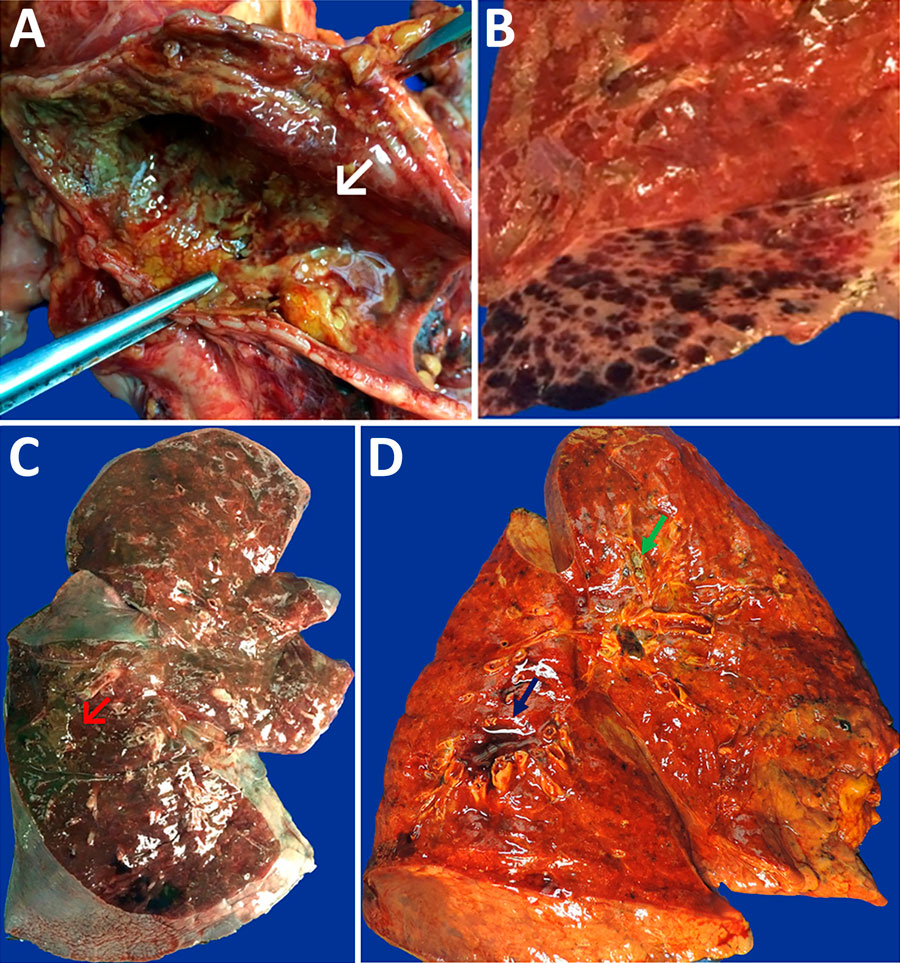
Figure 1. Macroscopic images of respiratory tract from patients with fatal yellow fever, São Paulo, Brazil, 2017–2019. A) Hemorrhagic necrosis of tracheal mucosa, covered with a thick whitish exudate (arrow), caused by Candida spp. invasive infection. B) Petechial pleural hemorrhage. C) Intense parenchymal edema and hemorrhage in the right lung, with massive gastrointestinal content aspiration in the posterior side (red arrow). D) Right lung with icterus, edema, hemorrhage, perivascular hemorrhage (blue arrow), and whitish exudate within bronchus (green arrow) caused by Aspergillus spp. infection.
Page created: January 20, 2026
Page updated: February 19, 2026
Page reviewed: February 19, 2026
The conclusions, findings, and opinions expressed by authors contributing to this journal do not necessarily reflect the official position of the U.S. Department of Health and Human Services, the Public Health Service, the Centers for Disease Control and Prevention, or the authors' affiliated institutions. Use of trade names is for identification only and does not imply endorsement by any of the groups named above.