Volume 32, Number 4—April 2026
Synopsis
Circulation Patterns, Genetic Diversity, and Public Health Implications of Enterovirus D68, Europe, 2014–2024
Cite This Article
Citation for Media
Abstract
Enterovirus D68 (EV-D68) represents a continuing public health concern, given its association with severe respiratory illness and neurologic complications. In this study, we analyzed EV-D68 circulation and genetic evolution during 2014–2024 using data from 18 countries in Europe. Of 61,297 enterovirus-positive specimens, molecular detection and viral protein 1 sequencing identified 3,541 (6%) EV-D68 cases. A biennial circulation pattern was observed; detection rates ranged from 9% in 2014 to 0.9% in 2019. The pattern was disrupted in 2020 because of measures implemented in response to the COVID-19 pandemic, but then notable increases occurred in 2021 (14%), 2022 (10.7%), and 2024 (20.6%). Subgenogroups B3 (59.8%) and A2/D (28.0%) were predominant; A2/D reemerged as dominant in 2024. Mutation analyses revealed changes in antigenic regions. Our findings underscore the persistent adaptation and resurgence of EV-D68 after COVID-19. Continued genomic surveillance is essential to monitor transmission patterns caused by antigenic changes.
Enterovirus D68 (EV-D68), a nonpolio enterovirus of the Picornaviridae family, has attracted attention because of its potential to cause severe respiratory illness and neurologic complications, particularly in children. First isolated in 1962, EV-D68 was sporadically detected until outbreaks in 2010 revealed its capacity for widespread transmission (1–4). Moreover, several clades (A–D) and subclades (A1, A2/D, B1, B2, B3, and C) have been described (5). Subsequent studies have also demonstrated that EV-D68 exhibits a cyclical pattern of circulation and a biennial recurrence (6,7), which continued until the COVID-19 pandemic, when the incidence of seasonal respiratory viruses was disrupted.
The ongoing circulation of EV-D68 poses a challenge for public health because outbreaks frequently coincide with increased rates of hospitalization for respiratory distress and, occasionally, cases of acute flaccid myelitis (AFM) (8). The neuropathogenic properties of the virus have been linked to specific amino acid residues. In particular, Leser et al. (9) recently identified 4 amino acid substitutions in the VP1 gene (I553L, D554N, A650T, and K835E) that are associated with neurovirulence in mice. Several recent studies have shown that neutralizing antibody responses to EV-D68 increase with age and that infection with 1 clade can generate cross-reactive immunity against other clades, providing key insights into population immunity and transmission dynamics (10–12).
Despite improvements in surveillance efforts, comprehensive analysis of EV-D68 circulation patterns and the elucidation of viral features remains limited. This study provides a summary of trends in EV-D68 circulation in Europe during 2014–2024, particularly focusing on prevalence, genetic diversity, and potential public health implications.
Data Collection
We obtained epidemiologic and molecular data from the passive and hospital-based surveillance systems (enterovirus, influenza-like illness, or acute flaccid paralysis [AFP]) of various institutions affiliated with the European Non-Poliovirus Enterovirus Network through a data collection form (Appendix Table 1) and other studies (1,3,13–16). Specimens from patients with symptoms suggestive of an enterovirus-related acute respiratory infection or neurologic illness (meningitis, AFP, or myelitis) were tested for virological confirmation according to case definition (17). Information regarding the samples that were tested and subsequently identified as EV-D68 was gathered during October 2014–December 2024. We also extracted demographics and clinical data from previous published works (1,3,13–16). Institutional Review Board approval (PR(AG)419/2023) was obtained from the Hospital Universitari Vall d’Hebron Clinical Research Ethics Committee.
Enterovirus Detection and Characterization
We conducted enterovirus detection using different real-time multiplex RT-PCRs (Table 1) (34). We subjected the partial viral protein (VP) 1 or the complete genome to sequencing for enterovirus typing (35) and genomic annotation of all enterovirus-positive specimens, as previously reported (1–3,33,36–38) (Table 1). We conducted further molecular characterization of EV-D68 by phylogenetic analyses using IQ-TREE multicore version 2.4.0 (Model Finder) (39) to define the genetic clades (Appendix Figure 1). References used for EV-D68 phylogeny at clade level are according to the following GenBank accession numbers: A1, KT959173; A2/D, F726085, KT959178, KU242683, and KY358058; B1, KP745751; B2, KP745768; B3, KT711083, KT803593, KU982558, and KY385886. In addition, we analyzed amino acid substitutions within antigenic epitopes of the VP1 protein, specifically the BC-loop (positions 642–655) and DE-loop (positions 692–698), and other areas recently linked to neuropathogenesis (9) using the Data Explorer module in MEGA6 (40). Numbering of amino acid positions in translated nucleotide sequences are relative to the Fermon strain (GenBank accession no. AY426531) and clinical isolate U.S./IL/14-18952 (GenBank accession no. KM851230) for the sequence annotation of immunogenicity and neuropathogenic sites. We determined the frequency of amino acid residues per antigenic site using EV-D68 A2/D and B3 clades, compared between prepandemic (2014–2019), pandemic (2020–2022), and postpandemic (2023–2024) periods, and plotted with Weblogo 3 (41). We compiled GenBank accession numbers of sequences used from the previously published studies and newly generated sequences (Appendix Table 2).
EV-D68 Circulation through Europe
Figure 1
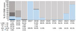
Figure 1. Yearly percentage and global distribution of EV-D68 cases, by clade, in study of circulation patterns, genetic diversity, and public health implications of EV-D68 circulation, Europe, 2014–2024. Numbers at bottom left...
During October 2014–December 2024, a total of 61,297 specimens from 50 institutions across 18 countries in Europe (Table 1) were laboratory-confirmed as enterovirus. A total of 3,541 (6%) of 61,297 were identified as EV-D68; of those, 2,339 (66%) were further genetically characterized based on the VP1 sequence (n = 2,057) or complete genome (n = 282). Subgenogroups B3 (1,398 [59.8%]) and A2/D (654 [28.0%]) were overall the most prevalent, followed by B2 (202 [8.6%]), B1 (71 [3.0%]), and A1 (14 [0.6%]) (Figure 1; Appendix Figure 1). EV-D68 was predominantly collected from persons with acute respiratory infection symptoms, primarily children; incidence among adolescents and adults (>15 years of age) increased during 2024 (Tables 2, 3).
Figure 2
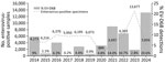
Figure 2. Yearly distribution of enterovirus-positive specimens and percentage of EV-D68–related infections in study of circulation patterns, genetic diversity, and public health implications of EV-D68 circulation, Europe, 2014–2024. EV-D68, enterovirus D68.
In Europe, EV-D68 circulation was predominantly observed during even years (9% in 2014, 6.3% in 2016, 4.4% in 2018), although circulation was disrupted in 2020 because of the implementation of preventive measures for SARS-CoV-2 (Figure 2). Nevertheless, we observed a notable upsurge in EV-D68 cases in 2021 (14.0%) across multiple countries (Figure 2). We also observed subsequent circulation in 2022 (10.7%) and during the 2024 season (20.6%). Although fewer cases were detected in 2023 (2.8%), the numbers were still substantially higher than in the low-circulation years before the pandemic (1.1% in 2015, 0.2% in 2017, and 0.9% in 2019) (Figure 2). Regardless of the changing epidemiology of EV-D68 over the years, we observed further levels of predominance of the EV-D68 clades over the study period (Figure 1). Although B3 prevailed in 2015 and 2016, A2/D dominated in 2017; both subgenogroups cocirculated in 2018. B3 was dominant again in the following years but decreased in 2024, when A2/D was the dominant subgenogroup (82.1%) (Figure 1).
Mutation Analyses of EV-D68 Strains
Immunogenic Epitopes
Figure 3
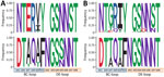
Figure 3. Frequency of amino acid changes within the antigenic epitopes of the viral protein 1 (BC- and DE-loops) in clades A2/D and B3 in study of circulation patterns, genetic diversity, and...
We have provided the frequency of amino acid changes detected at antigenic sites (BC- and DE-loops) of EV-D68 strains in prepandemic, pandemic, and postpandemic periods (Figure 3), as well as the complete mutation analysis of VP1 gene per year and per clade (Appendix Figures 2, 3). We observed a clear change in amino acid diversity in both A2/D (Appendix Figure 2) and B3 (Appendix Figure 3) clades during the 2 periods. Positions 647 and 650 in the BC-loop exhibited the greatest degree of variation compared with other sites in both clades (Figure 3). Within the A2/D clade, prepandemic sequences were predominantly characterized by glutamic acid at position 647 (E647) and valine at position 650 (V650). Although the isoleucine at position 650 (I650) dominated until 2016, a transition to valine (V650) was observed in 2017 and 2018; the detection rate during 2019 was 50% (Appendix Figure 2). Conversely, during the pandemic and postpandemic era (2020–2024), we observed a marked shift in residue predominance. Glycine at position 647 (G647) and isoleucine at position 650 (I650) became the dominant residues, largely replacing the prepandemic amino acids and indicating a substantial change in the antigenic landscape of the BC-loop (Figure 3; Appendix Figures 2, 4). Regarding the B3 clade, prepandemic sequences exhibited a high predominance of alanine at both position 647 (A647) and 650 (A650). In the aftermath of 2020, those residues underwent a substantial replacement by threonine at both positions (T647 and T650), which subsequently became predominant in postpandemic B3 sequences. In addition, within the DE-loop, position 695 exchange of amino acids before and after the pandemic (from S695 to N695) also indicates antigenic drift in this loop (Figure 3; Appendix Figures 3, 4).
Neurovirulent Epitopes
We further investigated the genetic diversity of the recently identified neurovirulence-associated markers within the VP1 gene (I553L, D554N, A650T, and K835E) (Table 4; Appendix Figure 4) (9). In the context of the A2/D clade, we observed no substantial changes among prepandemic, pandemic, and postpandemic periods; we observed neurovirulent amino acid changes at L553 and E835 and a glutamic acid at position 554 predominantly identified, in contrast to position 650, which was described previously as changing from a valine to an isoleucine (Table 4). With regard to the B3 clade, L553 was only identified in 2014, albeit in 1 sequence, whereas E554 was only identified in 2023–2024 in a shift from D to E. As described previously, we observed an apparent change to the neurovirulent threonine at position 650 (T650). In addition to A2/D, the neurovirulent glutamic acid at position 835 (E835) was detected throughout the decade (Table 4).
This study offers a comprehensive overview of the circulation of EV-D68 across Europe over a decade, emphasizing its prevalence, seasonality, and evolution. Our findings underscore the persistent adaptation and resurgence of EV-D68 in the postpandemic era.
Our analysis revealed a biennial seasonal pattern of EV-D68 activity and a pronounced prevalence during even years. That pattern is consistent with the findings of previous studies that have documented similar biennial trends in North America (42) and Europe (3,33,36,38). The interruption of the pattern in 2020 corroborates findings from other investigations that reported a reduced circulation of respiratory pathogens during the COVID-19 pandemic because of the implementation of preventive measures against SARS-CoV-2 (43). The resurgence of EV-D68 in 2021 and the subsequent peaks in 2022 and 2024 lend further support to the hypothesis that relaxing public health restrictions led to the reestablishment of EV-D68 transmission chains. Whether transmission reverts to a biennial cycle and changes in genetic diversity remain will require continued surveillance. Moreover, the predominance of EV-D68–related respiratory infections, especially in children, is cause for concern, particularly in the context of AFM. Although initial epidemiologic studies indicated a temporal association between EV-D68 outbreaks and AFM cases, subsequent neuropathological evidence demonstrating EV-D68 within anterior horn motor neurons has directly supported a causal relationship (2,44,45).
The high prevalence of EV-D68 in the 2024 season in some countries in Europe (20.6%) contrasts with earlier reports from the 2014 outbreak in Europe and United States, in which prevalence reached ≈9%–12% (19,37). That observation underscores the potential for variations in outbreak magnitude, which might be influenced by factors such as population immunity, viral evolution, public health interventions, and enhanced surveillance systems, particularly after the COVID-19 pandemic, which could have improved the detection of several respiratory viruses, such as EV-D68.
Genetic characterization of EV-D68 specimens revealed some replacements and dynamic genetic drifts in the predominance of subgenogroups B3 and A2/D over time, as has been reported in other regions and with similar yearly trends to this study (36,46,47). The exchange of clades might be associated with antigenic drift and immune selection pressures, which could have also driven the reemergence of A2/D as the dominant clade (82.1%) during 2024 with additional substitutions within the antigenic epitopes, as we observed. Moreover, that exchange could be related to differences in age distribution among B3- and A2/D-related cases, as previously described (36). During 2024, according to the available data, the ratio A2/D:B3 exhibited a higher prevalence among adults than in children (Table 2). That phenomenon could be attributed to immune evasion in adults, which is likely caused by changes in antigenic properties caused by mutations in antigenic epitopes, resulting in reduced neutralization by underlying neutralizing antibodies (36). Regarding that phenomenon, we observed amino acid changes within the VP1 gene, particularly in the BC-loop and DE-loop in positions 647, 650 (A2/D and B3), and 695 (B3), which mirror mutations reported during earlier outbreaks (36,47). Those changes could be associated with potential changes in viral fitness, neuropathogenesis, and immune evasion, as previously reported (9,48), or could represent an adaptive shift influencing receptor-binding properties and host-immune interactions (49). The significance of the identified changes, along with the changes seen over time in the BC-loop and DE-loop, require further investigations.
In addition to mutations in the VP1 region, changes outside the antigenic sites, such as the mice-neurovirulent E835 mutation, also deserve attention. E835 has been identified as a key factor in increasing neurotropism in mice, potentially promoting viral replication in motor neurons and enhancing retrograde axonal transport (9). In our series, E835 was mostly observed (instead of K835) in A2/D and B3. Also, the L553 mutation has been frequently reported in A2/D-related cases during the study period, which is also suspected to be a neurovirulence-related change in mice. Conversely, the B3 clade appears to have retained the isoleucine variant at this position, which is characterized as nonvirulent (15), yet we observed a shift to the neurovirulent threonine at position 650 after 2020. We observed a similar pattern at site 554, where there is a tendency to shift toward glutamic acid (E), another supposedly nonvirulent substitution seen in both the A2/D and B3 clades, although that residue position might also play a role in immune escape (15). The presumed neurovirulent genetic markers were identified in some A2/D- and B3-related cases in this study, but no increase in AFM-related cases was detected (8). Further research is required to ascertain whether those mutations or combinations of them are associated with severe clinical outcomes.
Of note, the emergence of SARS-CoV-2 coincided with increased mutational events on EV-D68, suggesting that competition with other respiratory pathogens during the pandemic might have driven such gradual changes (50). The circulation of new lineages that might have acquired an evolutionary advantage, possibly through immune evasion mechanisms (changes within antigenic epitopes), could then explain the predominance of the A2/D subgenogroup in 2024. Given the biennial epidemic cycles of EV-D68 and its apparent resurgence after the COVID-19 pandemic, global genomic monitoring is increasingly needed to identify emerging variants that could result in higher incidence and increased risk for neurologic complications. Resurgence shows an opportunity for targeted public health interventions, such as enhancing diagnostic capacity during anticipated peaks and promoting preventive measures.
Although this study provides valuable insights and is supported by both existing literature and recent findings, its first limitation is its reliance on laboratory-confirmed cases, which might potentially underestimate the true burden of EV-D68, because milder cases frequently go undiagnosed. In addition, although surveillance data might not be uniformly collected across Europe (and are largely unavailable at that level), the relevance of this study lies in the participation of 50 institutions from 18 countries in Europe. In addition, the limited number of fully genetically characterized cases might fail to capture the full diversity of circulating strains, at least during the 2024 season. Future studies should prioritize expanding genomic surveillance and integrating serologic data to better understand the effects of EV-D68 in both community and hospital populations, in relation to population-level immunity against this virus.
In conclusion, the reemergence of EV-D68 after the COVID-19 pandemic and its genetic evolution highlight current limitations that might pose a substantial public health challenge. An enhanced surveillance system based on real-time genomic monitoring of circulating viruses, coupled with a deeper understanding of the factors driving the evolution of this reemerging pathogen, is crucial to mitigating its impact.
Dr. Andrés is a postdoctoral researcher at Respiratory Viruses Unit in the Microbiology Department of Hospital Univeristari Vall d’Hebron. Her research interests are the epidemiology and molecular characterization of viruses, especially influenza viruses, enteroviruses, rhinoviruses, and coronaviruses.
Acknowledgments
We thank the European Society for Clinical Virology for hosting the European Non-Polio Enterovirus Network. We acknowledge all clinicians and technical staff participating in the European enterovirus/poliovirus surveillance programs in all participating laboratories and those who contributed to the collection, testing, and reporting of clinical and virological data presented in this study. We would specifically like to acknowledge Jeroen Cremer for sequencing all EV-D68 strains from the Netherlands and Lieuwe Roorda, Leo Smeets, and Thijs van de Laar for the additional 2024 EV-D68 sequence data from the Netherlands to complete the data collected over the study period.
This research was partially supported by Fondo de Investigación Sanitaria, Ministerio Español de Economía y Competitividad (grant no. FIS PI22/00023) and CIBER–Consorcio Centro de Investigación Biomédica en Red (CIBERINFEC, ISCIII–CIBER de Enfermedades Infecciosas), Instituto de Salud Carlos III, Ministerio de Ciencia e Innovación, and Unión Europea–NextGenerationEU.
During the preparation of this work, the authors used ChatGPT to improve readability and clarity. After using this tool, all authors reviewed and edited the content as needed and took full responsibility for the content of the publication.
References
- Benschop KS, Albert J, Anton A, Andrés C, Aranzamendi M, Armannsdóttir B, et al. Re-emergence of enterovirus D68 in Europe after easing the COVID-19 lockdown, September 2021. Euro Surveill. 2021;26:
2100998 . DOIPubMedGoogle Scholar - Simoes MP, Hodcroft EB, Simmonds P, Albert J, Alidjinou EK, Ambert-Balay K, et al. Epidemiological and clinical insights into the enterovirus D68 upsurge in Europe 2021–2022 and emergence of novel B3-derived lineages, ENPEN Multicentre Study. J Infect Dis. 2024;230:e917–28. DOIPubMedGoogle Scholar
- Andrés C, Vila J, Creus-Costa A, Piñana M, González-Sánchez A, Esperalba J, et al. Enterovirus D68 in hospitalized children, Barcelona, Spain, 2014–2021. Emerg Infect Dis. 2022;28:1327–31. DOIPubMedGoogle Scholar
- Messacar K, Tyler KL. Enterovirus D68-associated acute flaccid myelitis: rising to the clinical and research challenges. JAMA. 2019;321:831–2. DOIPubMedGoogle Scholar
- Tokarz R, Firth C, Madhi SA, Howie SRC, Wu W, Sall AA, et al. Worldwide emergence of multiple clades of enterovirus 68. J Gen Virol. 2012;93:1952–8. DOIPubMedGoogle Scholar
- Messacar K, Pretty K, Reno S, Dominguez SR. Continued biennial circulation of enterovirus D68 in Colorado. J Clin Virol. 2019;113:24–6. DOIPubMedGoogle Scholar
- Andrés C, Vila J, Gimferrer L, Piñana M, Esperalba J, Codina MG, et al. Surveillance of enteroviruses from paediatric patients attended at a tertiary hospital in Catalonia from 2014 to 2017. J Clin Virol. 2019;110:29–35. DOIPubMedGoogle Scholar
- Helfferich J, Calvo C, Alpeter E, Andrés C, Antón A, Aubart M, et al. Acute flaccid myelitis in Europe between 2016 and 2023: indicating the need for better registration. Euro Surveill. 2025;30:
2400579 . DOIPubMedGoogle Scholar - Leser JS, Frost JL, Wilson CJ, Rudy MJ, Clarke P, Tyler KL. VP1 is the primary determinant of neuropathogenesis in a mouse model of enterovirus D68 acute flaccid myelitis. J Virol. 2024;98:
e0039724 . DOIPubMedGoogle Scholar - Pons-Salort M, Lambert B, Kamau E, Pebody R, Harvala H, Simmonds P, et al. Changes in transmission of enterovirus D68 (EV-D68) in England inferred from seroprevalence data. eLife. 2023;12:12. DOIPubMedGoogle Scholar
- Kamau E, Harvala H, Blomqvist S, Nguyen D, Horby P, Pebody R, et al. Increase in enterovirus D68 infections in young children, United Kingdom, 2006–2016. Emerg Infect Dis. 2019;25:1200–3. DOIPubMedGoogle Scholar
- Karelehto E, Koen G, Benschop K, van der Klis F, Pajkrt D, Wolthers K. Enterovirus D68 serosurvey: evidence for endemic circulation in the Netherlands, 2006 to 2016. Euro Surveill. 2019;24:
1800671 . DOIPubMedGoogle Scholar - Poelman R, Schuffenecker I, Van Leer-Buter C, Josset L, Niesters HG, Lina B. ESCV-ECDC EV-D68 study group. European surveillance for enterovirus D68 during the emerging North-American outbreak in 2014. J Clin Virol. 2015;71:1–9. DOIPubMedGoogle Scholar
- de Schrijver S, Vanhulle E, Ingenbleek A, Alexakis L, Johannesen CK, Broberg EK, et al.; ENPEN Study Collaborators. Epidemiological and clinical insights into enterovirus circulation in Europe, 2018–2023: a multi-center retrospective surveillance study. J Infect Dis. 2025;232:e104–15. DOIPubMedGoogle Scholar
- Hirvonen A, Johannesen CK, Simmonds P, Fischer TK, Harvala H, Benschop KSM, et al.; ENPEN study collaborators. Sustained circulation of enterovirus D68 in Europe in 2023 and the continued evolution of enterovirus D68 B3-lineages associated with distinct amino acid substitutions in VP1 protein. J Clin Virol. 2025;178:
105785 . DOIPubMedGoogle Scholar - Bubba L, Broberg EK, Jasir A, Simmonds P, Harvala H, Redlberger-Fritz M, et al.; Enterovirus study collaborators. Circulation of non-polio enteroviruses in 24 EU and EEA countries between 2015 and 2017: a retrospective surveillance study. Lancet Infect Dis. 2020;20:350–61. DOIPubMedGoogle Scholar
- Harvala H, Benschop KSM, Berginc N, Midgley S, Wolthers K, Simmonds P, et al.; On Behalf Of The Enpen Hospital-Based Surveillance Network. European Non-Polio Enterovirus Network: introduction of hospital-based surveillance network to understand the true disease burden of non-polio enterovirus and parechovirus infections in Europe. Microorganisms. 2021;9:1827. DOIPubMedGoogle Scholar
- Nix WA, Oberste MS, Pallansch MA. Sensitive, seminested PCR amplification of VP1 sequences for direct identification of all enterovirus serotypes from original clinical specimens. J Clin Microbiol. 2006;44:2698–704. DOIPubMedGoogle Scholar
- Midgley CM, Watson JT, Nix WA, Curns AT, Rogers SL, Brown BA, et al. EV-D68 Working Group. Severe respiratory illness associated with a nationwide outbreak of enterovirus D68 in the USA (2014): a descriptive epidemiological investigation. Lancet Respir Med. 2015;3:879–87. DOIPubMedGoogle Scholar
- Schuffenecker I, Mirand A, Josset L, Henquell C, Hecquet D, Pilorgé L, et al. Epidemiological and clinical characteristics of patients infected with enterovirus D68, France, July to December 2014. Euro Surveill. 2016;21. DOIPubMedGoogle Scholar
- Keeren K, Böttcher S, Diedrich S. Enterovirus Surveillance (EVSurv) in Germany. Microorganisms. 2021;9:2005. DOIPubMedGoogle Scholar
- Diedrich S, Driesel G, Schreier E. Sequence comparison of echovirus type 30 isolates to other enteroviruses in the 5′ noncoding region. J Med Virol. 1995;46:148–52. DOIPubMedGoogle Scholar
- Dierssen U, Rehren F, Henke-Gendo C, Harste G, Heim A. Rapid routine detection of enterovirus RNA in cerebrospinal fluid by a one-step real-time RT-PCR assay. J Clin Virol. 2008;42:58–64. DOIPubMedGoogle Scholar
- Bragstad K, Jakobsen K, Rojahn AE, Skram MK, Vainio K, Holberg-Petersen M, et al. High frequency of enterovirus D68 in children hospitalised with respiratory illness in Norway, autumn 2014. Influenza Other Respir Viruses. 2015;9:59–63. DOIPubMedGoogle Scholar
- Oberste MS, Maher K, Kilpatrick DR, Flemister MR, Brown BA, Pallansch MA. Typing of human enteroviruses by partial sequencing of VP1. J Clin Microbiol. 1999;37:1288–93. DOIPubMedGoogle Scholar
- Oberste MS, Maher K, Kilpatrick DR, Pallansch MA. Molecular evolution of the human enteroviruses: correlation of serotype with VP1 sequence and application to picornavirus classification. J Virol. 1999;73:1941–8. DOIPubMedGoogle Scholar
- Piralla A, Girello A, Grignani M, Gozalo-Margüello M, Marchi A, Marseglia G, et al. Phylogenetic characterization of enterovirus 68 strains in patients with respiratory syndromes in Italy. J Med Virol. 2014;86:1590–3. DOIPubMedGoogle Scholar
- Piralla A, Girello A, Premoli M, Baldanti F. A new real-time reverse transcription-PCR assay for detection of human enterovirus 68 in respiratory samples. J Clin Microbiol. 2015;53:1725–6. DOIPubMedGoogle Scholar
- Benschop K, Molenkamp R, van der Ham A, Wolthers K, Beld M. Rapid detection of human parechoviruses in clinical samples by real-time PCR. J Clin Virol. 2008;41:69–74. DOIPubMedGoogle Scholar
- Jaramillo-Gutierrez G, Benschop KS, Claas EC, de Jong AS, van Loon AM, Pas SD, et al. September through October 2010 multi-centre study in the Netherlands examining laboratory ability to detect enterovirus 68, an emerging respiratory pathogen. J Virol Methods. 2013;190:53–62. DOIPubMedGoogle Scholar
- Hayes A, Nguyen D, Andersson M, Antón A, Bailly JL, Beard S, et al. A European multicentre evaluation of detection and typing methods for human enteroviruses and parechoviruses using RNA transcripts. J Med Virol. 2020;92:1065–74. DOIPubMedGoogle Scholar
- Cabrerizo M, Echevarria JE, González I, de Miguel T, Trallero G. Molecular epidemiological study of HEV-B enteroviruses involved in the increase in meningitis cases occurred in Spain during 2006. J Med Virol. 2008;80:1018–24. DOIPubMedGoogle Scholar
- González-Sanz R, Taravillo I, Reina J, Navascués A, Moreno-Docón A, Aranzamendi M, et al. Enterovirus D68-associated respiratory and neurological illness in Spain, 2014–2018. Emerg Microbes Infect. 2019;8:1438–44. DOIPubMedGoogle Scholar
- Harvala H, Jasir A, Penttinen P, Pastore Celentano L, Greco D, Broberg E. Surveillance and laboratory detection for non-polio enteroviruses in the European Union/European Economic Area, 2016. Euro Surveill. 2017;22:16–00807. DOIPubMedGoogle Scholar
- Kroneman A, Vennema H, Deforche K, v d Avoort H, Peñaranda S, Oberste MS, et al. An automated genotyping tool for enteroviruses and noroviruses. J Clin Virol. 2011;51:121–5. DOIPubMedGoogle Scholar
- Hodcroft EB, Dyrdak R, Andrés C, Egli A, Reist J, García Martínez de Artola D, et al. Evolution, geographic spreading, and demographic distribution of enterovirus D68. PLoS Pathog. 2022;18:
e1010515 . DOIPubMedGoogle Scholar - Bal A, Sabatier M, Wirth T, Coste-Burel M, Lazrek M, Stefic K, et al. Emergence of enterovirus D68 clade D1, France, August to November 2018. Euro Surveill. 2019;24:
1800699 . DOIPubMedGoogle Scholar - Pellegrinelli L, Giardina F, Lunghi G, Uceda Renteria SC, Greco L, Fratini A, et al. Emergence of divergent enterovirus (EV) D68 sub-clade D1 strains, northern Italy, September to October 2018. Euro Surveill. 2019;24:
1900090 . DOIPubMedGoogle Scholar - Kalyaanamoorthy S, Minh BQ, Wong TKF, von Haeseler A, Jermiin LS. ModelFinder: fast model selection for accurate phylogenetic estimates. Nat Methods. 2017;14:587–9. DOIPubMedGoogle Scholar
- Tamura K, Peterson D, Peterson N, Stecher G, Nei M, Kumar S. MEGA5: molecular evolutionary genetics analysis using maximum likelihood, evolutionary distance, and maximum parsimony methods. Mol Biol Evol. 2011;28:2731–9. DOIPubMedGoogle Scholar
- Crooks GE, Hon G, Chandonia JM, Brenner SE. WebLogo: a sequence logo generator. Genome Res. 2004;14:1188–90. DOIPubMedGoogle Scholar
- Messacar K, Abzug MJ, Dominguez SR. The emergence of enterovirus-D68. Microbiol Spectr. 2016;4:4.3.37. DOIPubMedGoogle Scholar
- Britton PN, Hu N, Saravanos G, Shrapnel J, Davis J, Snelling T, et al. COVID-19 public health measures and respiratory syncytial virus. Lancet Child Adolesc Health. 2020;4:e42–3. DOIPubMedGoogle Scholar
- Messacar K, Asturias EJ, Hixon AM, Van Leer-Buter C, Niesters HGM, Tyler KL, et al. Enterovirus D68 and acute flaccid myelitis—evaluating the evidence for causality. Lancet Infect Dis. 2018;18:e239–47. DOIPubMedGoogle Scholar
- Vogt MR, Wright PF, Hickey WF, De Buysscher T, Boyd KL, Crowe JE Jr. Enterovirus D68 in the anterior horn cells of a child with acute flaccid myelitis. N Engl J Med. 2022;386:2059–60. DOIPubMedGoogle Scholar
- Wang H, Diaz A, Moyer K, Mele-Casas M, Ara-Montojo MF, Torrus I, et al. Molecular and clinical comparison of enterovirus D68 outbreaks among hospitalized children, Ohio, USA, 2014 and 2018. Emerg Infect Dis. 2019;25:2055–63. DOIPubMedGoogle Scholar
- Shi Y, Liu Y, Wu Y, Hu S, Sun B. Molecular epidemiology and recombination of enterovirus D68 in China. Infect Genet Evol. 2023;115:
105512 . DOIPubMedGoogle Scholar - Brown DM, Hixon AM, Oldfield LM, Zhang Y, Novotny M, Wang W, et al. Contemporary circulating enterovirus D68 strains have acquired the capacity for viral entry and replication in human neuronal cells. MBio. 2018;9:e01954–18. DOIPubMedGoogle Scholar
- Fall A, Abdullah O, Han L, Norton JM, Gallagher N, Forman M, et al. Enterovirus D68: genomic and clinical comparison of 2 seasons of increased viral circulation and discrepant incidence of acute flaccid myelitis—Maryland, USA. Open Forum Infect Dis. 2024;11:
ofae656 . DOIPubMedGoogle Scholar - Dhanasekaran V, Sullivan S, Edwards KM, Xie R, Khvorov A, Valkenburg SA, et al. Human seasonal influenza under COVID-19 and the potential consequences of influenza lineage elimination. Nat Commun. 2022;13:1721. DOIPubMedGoogle Scholar
Figures
Tables
Cite This ArticleOriginal Publication Date: April 07, 2026
Table of Contents – Volume 32, Number 4—April 2026
| EID Search Options |
|---|
|
|
|
|
|
|
Please use the form below to submit correspondence to the authors or contact them at the following address:
Cristina Andrés, Respiratory Viruses Unit—Virology Section, Microbiology Department, Hospital Universitari Vall d’Hebron, Passeig Vall d’Hebron 119-129, 08035 Barcelona, Spain
Top