Yellow Fever Virus Surveillance in Callithrix spp. Marmosets during Epizootic Outbreak, Brazil, 2024–2025
Márcio Junio Lima Siconelli, Jéssica Caroline de Almeida Dias, Eduardo Ferreira Machado, Mariana Sequetin Cunha, Natália Coelho Couto de Azevedo Fernandes, Juliana Mariotti Guerra, Luana Bonon, Alline Borges Salomão, Karin Werther, Karina Paes Bürger, Adolorata Aparecida Bianco Carvalho, Daniel Marques, and Benedito Antonio Lopes da Fonseca

Author affiliation: Unidade de Vigilância de Zoonoses da Divisão de Vigilância Ambiental em Saúde, Secretaria Municipal da Saúde, Ribeirão Preto, Brazil (M.J.L. Siconelli); Faculdade de Medicina de Ribeirão Preto, Universidade de São Paulo, Ribeirão Preto (M.J.L. Siconelli, J.C. de Almeida Dias, B.A. Lopes da Fonseca); Centro de Patologia, Instituto Adolfo Lutz de São Paulo, Secretaria Estadual da Saúde de São Paulo, São Paulo, Brazil (E.F. Machado, N.C.C.A. Fernandes); Centro de Virologia, Instituto Adolfo Lutz de São Paulo, Secretaria Estadual da Saúde de São Paulo, São Paulo (M.S. Cunha); Universidade de São Paulo Departamento de Patologia da Faculdade de Medicina Veterinária e Zootecnia, São Paulo (J.M. Guerra); Programa de Residência em Área Profissional da Saúde, Medicina Veterinária e Saúde, do Departamento de Patologia, Reprodução e Saúde Única da Faculdade de Ciências Agrárias e Veterinárias, Universidade Estadual Paulista, Jaboticabal, Brazil (L. Bonon, A.B. Salomão, K. Werther, K.P. Bürger, A.A.B. Carvalho); Grupo de Vigilância Epidemiológica, Centro de Vigilância Epidemiológica “Prof. Alexandre Vranjac,” Secretaria Estadual da Saúde de São Paulo, São Paulo (D. Marques)
Main Article
Figure 2
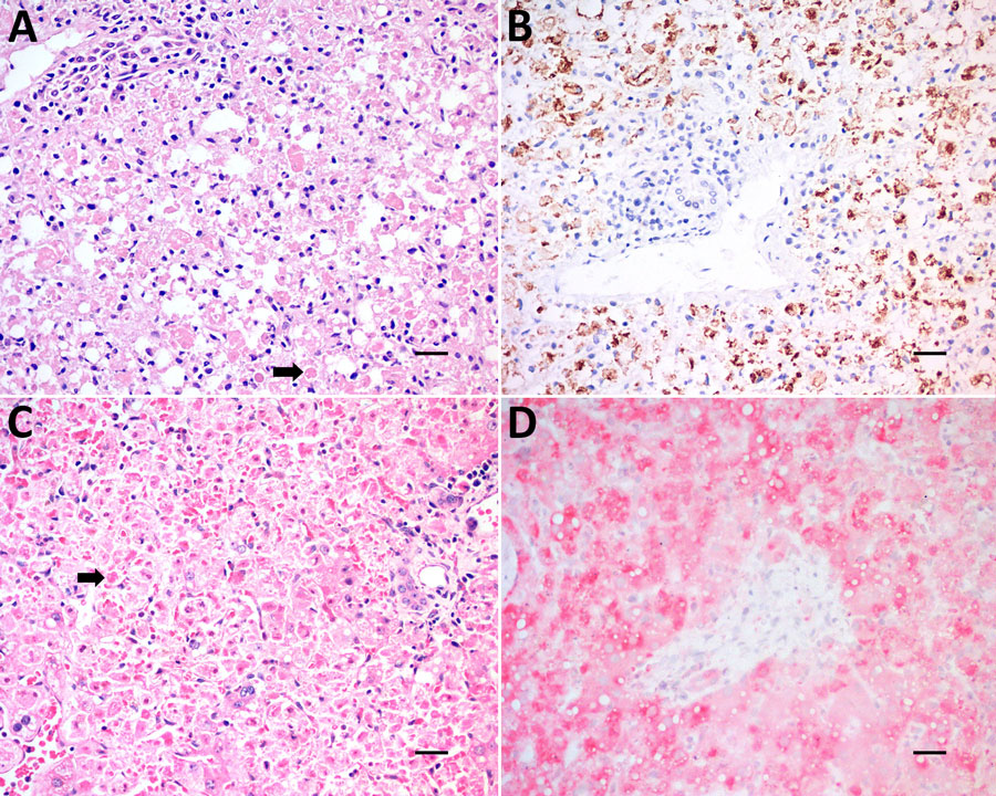
Figure 2. Laboratory analysis of liver samples from nonhuman primates collected for yellow fever virus (YFV) surveillance in Callithrix spp. marmosets during epizootic outbreak, Brazil, 2024–2025. A, B) Samples from Callicebus nigrifrons black fronted titi monkeys; C, D) samples from Alouatta caraya black howler monkeys. A) Hematoxylin and eosin stain of liver showing acute and severe hepatic damage characterized by diffusely individual cellular apoptosis and necrosis with Councilman–Rocha Lima bodies (arrow). B) Immunohistochemistry stain of hepatocytes; brown stain indicates cells positive for YFV antigen. C) Immunohistochemistry stain showing acute and severe hepatic damage characterized by individual cellular apoptosis and necrosis with Councilman–Rocha Lima bodies (arrow); brown indicates hepatocytes positive by YFV antigen. D) Immunohistochemistry stain of hepatocytes positive for YFV antigen (red). Scale bars indicate 20 µm.
Main Article
Page created: April 17, 2026
Page updated: June 01, 2026
Page reviewed: June 01, 2026
The conclusions, findings, and opinions expressed by authors contributing to this journal do not necessarily reflect the official position of the U.S. Department of Health and Human Services, the Public Health Service, the Centers for Disease Control and Prevention, or the authors' affiliated institutions. Use of trade names is for identification only and does not imply endorsement by any of the groups named above.