Volume 32, Number 6—June 2026
Dispatch
Placental Vascular Pathology Associated with Congenital Lymphocytic Choriomeningitis Virus Infection, Philadelphia, Pennsylvania, USA
Abin Abraham, Rebecca L. Linn, Dustin D. Flannery, and Scott M. Gordon
Figure 2
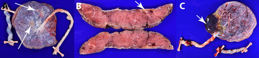
Figure 2. Pathologic findings of placental vascular anomalies associated with congenital lymphocytic choriomeningitis virus infection, Philadelphia, Pennsylvania, USA. A, B) Placenta from case 1; C) placenta from case 2. A) Gross pathology of the fetal surface showing yellow-white subchorionic intervillous thrombi (arrows). B) Cross sections of the placenta showing a subchorionic intervillous thrombus (arrow) and a peripheral intraparenchymal thrombus (white dashed circle). C) Gross pathology of the fetal surface of placenta with marginal insertion of three vessel umbilical cord and acute subamniotic hemorrhage (arrow). No additional gross lesions were identified after serial sectioning.
Page created: April 23, 2026
Page updated: June 01, 2026
Page reviewed: June 01, 2026
The conclusions, findings, and opinions expressed by authors contributing to this journal do not necessarily reflect the official position of the U.S. Department of Health and Human Services, the Public Health Service, the Centers for Disease Control and Prevention, or the authors' affiliated institutions. Use of trade names is for identification only and does not imply endorsement by any of the groups named above.