Volume 32, Number 6—June 2026
Dispatch
Placental Vascular Pathology Associated with Congenital Lymphocytic Choriomeningitis Virus Infection, Philadelphia, Pennsylvania, USA
Figure 3
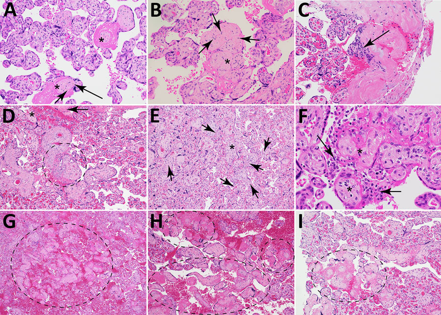
Figure 3. Microscopic findings for placental vascular pathology associated with congenital lymphocytic choriomeningitis virus infection, Philadelphia, Pennsylvania, USA. Hematoxylin and eosin–stained cross-sections of placenta from case 1 (A, B) and case 2 (C–I). A) Avascular villi surrounded by pink perivillous fibrin (asterisks) with scattered foci of rust-colored hemosiderin deposition (arrows) within the hyalinized stroma. Original magnification ×200. B) Cluster of avascular villi in the center of the image (asterisk) with scattered intravillous lymphocytes (arrows), compatible with chronic villitis. Original magnification ×200. C) Plasma cells (arrow) within the basal plate, consistent with chronic deciduitis. Original magnification ×200. D) Chronic villitis and perivillitis involving stem villi vessels (circle), with an associated avascular villus (asterisk) and fibrin deposition (arrow). Original magnification ×100. E) Chronic villitis (asterisk) with villous stromal vascular karyorrhexis; arrows point to examples of extravasated, fragmented erythrocytes and nuclear debris. Original magnification ×100. F) Histiocytic intervillositis (arrows) adjacent to avascular villi (asterisks) with perivillous fibrin deposition. Original magnification ×400. G–I) Avascular villi with associated perivillous fibrin deposition (dashed ellipses). G) Original magnification ×40; H, I) original magnification ×100.