Skip directly to site content Skip directly to page options Skip directly to A-Z link Skip directly to A-Z link Skip directly to A-Z link

Disclaimer: Early release articles are not considered as final versions. Any changes will be reflected in the online version in the month the article is officially released.

Volume 32, Number 7—July 2026

Research Letter

Ophthalmomyiasis Outbreak Caused by Oestrus ovis Infection, Algeria, 2025

Yuyang Zeng1, Hongkuan Yang1, Xin Li, Hongzheng Yang, and Yunyun ZhouComments to Author 
Author affiliation: Byers Eye Institute at Stanford University School of Medicine, Palo Alto, California, USA (Y. Zeng); Renmin Hospital of Wuhan University, Wuhan, China (Y. Zeng, Y. Zhou); Huazhong University of Science and Technology, Wuhan (Hongkuan Yang, X. Li); Renhe Hospital Affiliated with People’s Republic of China Three Gorges University, Yichang, China (Hongzheng Yang).

Main Article

Figure 1

Clinical ocular findings in patients with acute external ophthalmomyiasis caused by Oestrus ovis infection after sheep exposure during Eid al-Adha, Algeria, 2025. A–C) Representative external ocular photographs show acute conjunctivitis-like findings, including palpebral edema, conjunctival hyperemia, and mucous discharge. D, E) Slit-lamp examination images show motile larvae on the corneal surface and in the lower conjunctival fornix. F–I) Slit-lamp examination images show conjunctival inflammation, including conjunctival congestion, edema, and mucous discharge. Black arrow in panel E indicates the location of an O. ovis larva.

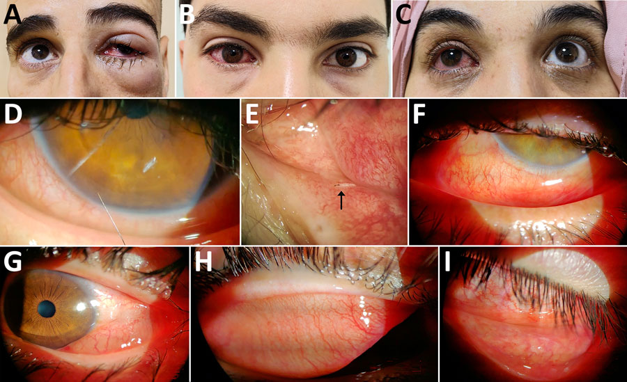
Figure 1. Clinical ocular findings in patients with acute external ophthalmomyiasis caused by Oestrus ovis infection after sheep exposure during Eid al-Adha, Algeria, 2025. A–C) Representative external ocular photographs show acute conjunctivitis-like findings, including palpebral edema, conjunctival hyperemia, and mucous discharge. D, E) Slit-lamp examination images show motile larvae on the corneal surface and in the lower conjunctival fornix. F–I) Slit-lamp examination images show conjunctival inflammation, including conjunctival congestion, edema, and mucous discharge. Black arrow in panel E indicates the location of an O. ovis larva.

Main Article

1These first authors contributed equally to this article.

Page created: May 28, 2026
Page updated: June 24, 2026
Page reviewed: June 24, 2026
The conclusions, findings, and opinions expressed by authors contributing to this journal do not necessarily reflect the official position of the U.S. Department of Health and Human Services, the Public Health Service, the Centers for Disease Control and Prevention, or the authors' affiliated institutions. Use of trade names is for identification only and does not imply endorsement by any of the groups named above.
file_external