Volume 9, Number 3—March 2003
Synopsis
Electron Microscopy for Rapid Diagnosis of Emerging Infectious Agents1
Figure 1
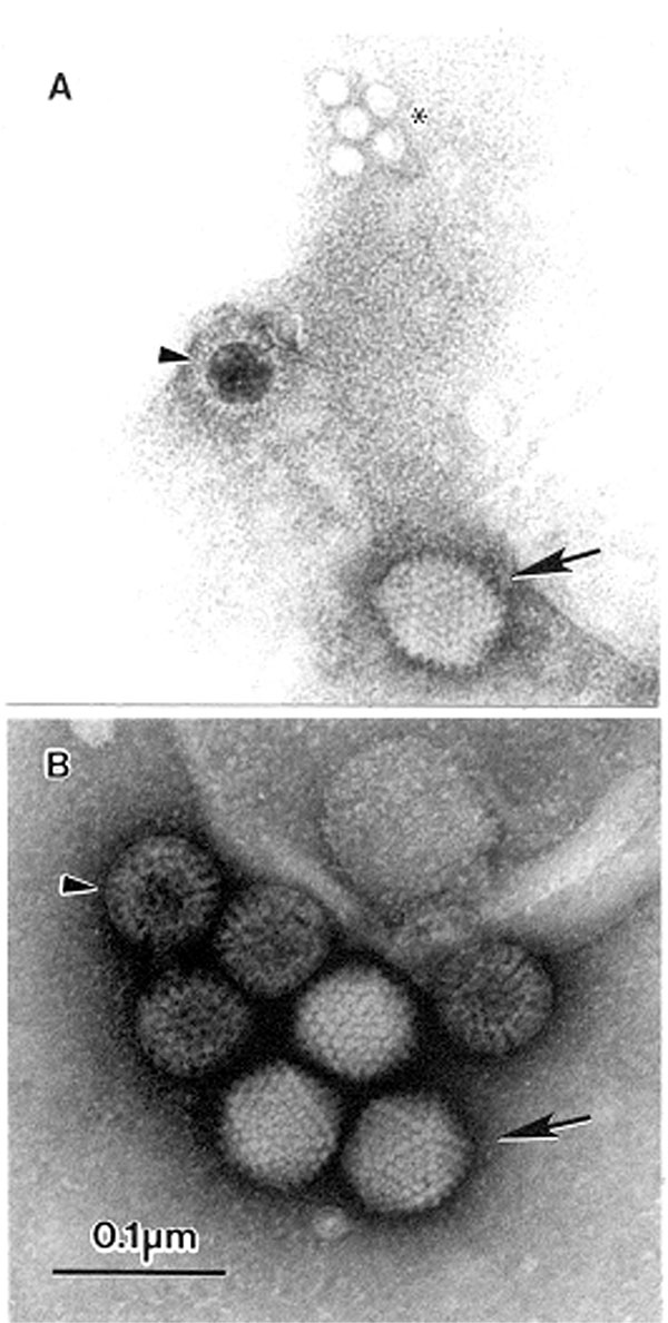
Figure 1. The open view of diagnostic electron microscopyAMultiple agents observed in a fecal sample from a pediatric patient with diarrheaA 10% suspension was prepared in distilled water, cleared by low-speed centrifugation followed by 5 minutes at 15,000 x g in a bench top centrifuge, and centrifuged directly to the grid using an Airfuge EM-90 rotor (Beckman, Palo Alto, CA): adenovirus-(→), incomplete rotavirus-particle (>), and small round featureless particles, probably adeno-associated virus (ρ) phosphotungstic acid stainedBDouble infection with adenovirus (→) and complete rotavirus particles (>), in the stool of a 1-yearold childThe sample was suspended 1:3 in distilled water, cleared by low-speed centrifugation, and prepared for examination by the two-step methodAqueous uranyl acetate stainedBar = 100 nm.