Skip directly to site content Skip directly to page options Skip directly to A-Z link Skip directly to A-Z link Skip directly to A-Z link
Volume 9, Number 3—March 2003
Synopsis

Electron Microscopy for Rapid Diagnosis of Emerging Infectious Agents1

Paul R. Hazelton*Comments to Author  and Hans R. Gelderblom†
Author affiliations: *University of Manitoba, Winnipeg, MB, Canada; †Robert Koch-Institut, Berlin, Germany

Main Article

Figure 5

Comparison of herpesvirus appearance after positive and negative stain electron microscopic. A. Positive staining. Samples undergo a lengthy process of fixation, incubation with heavy metal ions (osmium, uranyl), dehydration, embedment, ultrathin sectioning, and staining. Chemical moieties in the object show differential affinities for the heavy metal stains, resulting in a clear outline of the viral bilayer envelope, viral envelope proteins, nucleocapsid, and the dense nucleic acid containing c

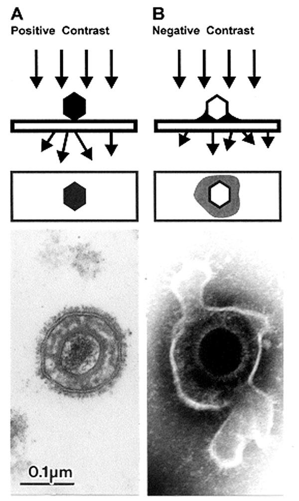
Figure 5. Comparison of herpesvirus appearance after positive and negative stain electron microscopicAPositive stainingSamples undergo a lengthy process of fixation, incubation with heavy metal ions (osmium, uranyl), dehydration, embedment, ultrathin sectioning, and stainingChemical moieties in the object show differential affinities for the heavy metal stains, resulting in a clear outline of the viral bilayer envelope, viral envelope proteins, nucleocapsid, and the dense nucleic acid containing coreBNegative stainingAfter a brief fixation, samples are mounted directly on electron microscopic grids and stained as in Figure 4The electron-dense stain (phosphotungstic acid [phosphotungstic acid], uranyl acetate, and the like) penetrates the virion and embeds the particle in a matrix of stainDue to density differences between the stain and weakly scattering biological components of the virion, the virion appears as a transparent and detailed reverse (negative) imagePenetration of stain into the nucleocapsid provides a dense core with the crenellated appearance presented by the central channel of capsomers on the nucleocapsid surfaceViral surface proteins appear as projections from the labile envelopephosphotungstic acidBar = 100 nm.

Main Article

1Both authors contributed equally to this review.

Page created: December 07, 2010
Page updated: December 07, 2010
Page reviewed: December 07, 2010
The conclusions, findings, and opinions expressed by authors contributing to this journal do not necessarily reflect the official position of the U.S. Department of Health and Human Services, the Public Health Service, the Centers for Disease Control and Prevention, or the authors' affiliated institutions. Use of trade names is for identification only and does not imply endorsement by any of the groups named above.
file_external