Volume 9, Number 3—March 2003
Synopsis
Electron Microscopy for Rapid Diagnosis of Emerging Infectious Agents1
Paul R. Hazelton* and Hans R. Gelderblom†
Figure 3
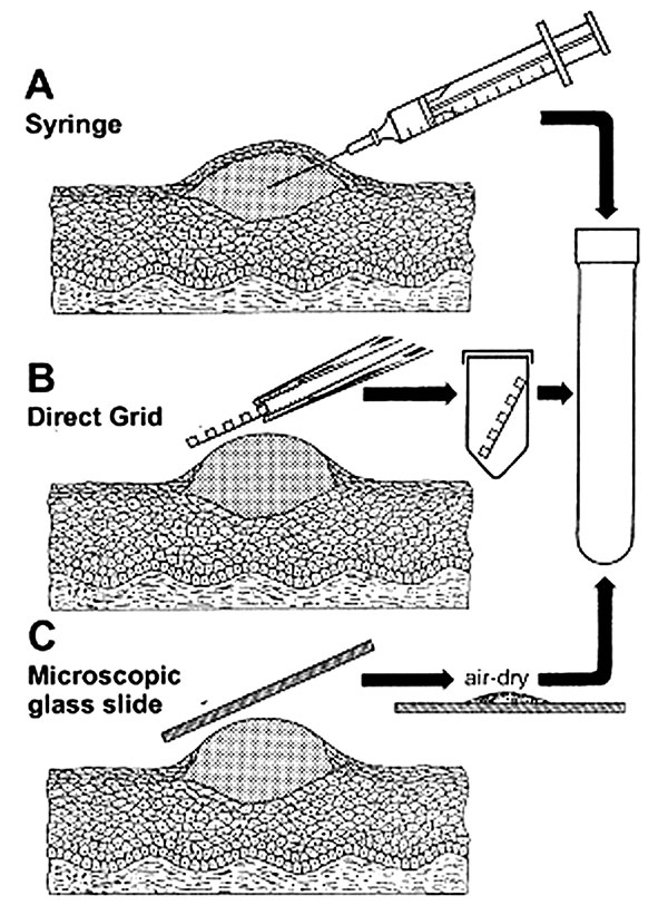
Figure 3. Three methods for efficient collection of vesicular and blister fluids for diagnostic electron microscopicAThe contents of a vesicle are collected into the barrel of a needleBAfter the blister is opened, a coated electron microscopic grid is touched to the fluid and air-dried (direct electron microscopic)CA glass microscope slide is touched directly to an unroofed lesion and a smear preparedSamples are then placed in rigid containers for transport to the electron microscopic laboratory.
Page created: December 07, 2010
Page updated: December 07, 2010
Page reviewed: December 07, 2010
The conclusions, findings, and opinions expressed by authors contributing to this journal do not necessarily reflect the official position of the U.S. Department of Health and Human Services, the Public Health Service, the Centers for Disease Control and Prevention, or the authors' affiliated institutions. Use of trade names is for identification only and does not imply endorsement by any of the groups named above.