Volume 14, Number 9—September 2008
Research
Circulation of 3 Lineages of a Novel Saffold Cardiovirus in Humans
Ana Maria Bispo de Filippis, Luciano Kleber de Souza Luna, Andreas Stöcker, Patrícia Silva Almeida, Tereza Cristina Medrado Ribeiro, Nadine Petersen, Petra Herzog, Célia Pedroso, Hans Iko Huppertz, Hugo da Costa Ribeiro, Sigrid Baumgarte, and Sung Sup Park
Figure 1
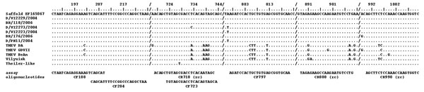
Figure 1. Nucleic acid alignment of the hybridization sites of diagnostic reverse transcription–PCR oligonucleotides. Oligonucleotides are shown below the alignment panel. The base count in the top line is based on Saffold virus, which also serves as the comparison sequence in the alignment. Dots represent identical bases in compared sequences; deviations are spelled out. A slash (/) represents a gap in the alignment; (rc) means that the reverse complementary sequence is shown for the antisense primer.
Page created: July 13, 2010
Page updated: July 13, 2010
Page reviewed: July 13, 2010
The conclusions, findings, and opinions expressed by authors contributing to this journal do not necessarily reflect the official position of the U.S. Department of Health and Human Services, the Public Health Service, the Centers for Disease Control and Prevention, or the authors' affiliated institutions. Use of trade names is for identification only and does not imply endorsement by any of the groups named above.