Volume 2, Number 3—July 1996
Synopsis
Application of Molecular Techniques to the Diagnosis of Microsporidial Infection
Daniel P. Fedorko and Yasmine M. Hijazi
Figure 3
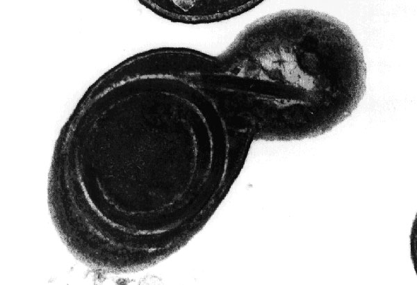
Figure 3. Transmission electron micrograph of a cell culture-derived Encephalitozoon intestinalis spore showing the polar tubule in the process of being extruded. The coiled arrangement of the tubule within the spore is clearly demonstrated. Original magnification, x39,000.
Page created: December 20, 2010
Page updated: December 20, 2010
Page reviewed: December 20, 2010
The conclusions, findings, and opinions expressed by authors contributing to this journal do not necessarily reflect the official position of the U.S. Department of Health and Human Services, the Public Health Service, the Centers for Disease Control and Prevention, or the authors' affiliated institutions. Use of trade names is for identification only and does not imply endorsement by any of the groups named above.