Volume 20, Number 12—December 2014
Research
Replication and Shedding of MERS-CoV in Upper Respiratory Tract of Inoculated Dromedary Camels
Danielle R. Adney, Neeltje van Doremalen, Vienna R. Brown, Trenton Bushmaker, Dana Scott, Emmie de Wit, Richard A. Bowen1 , and Vincent J. Munster1
Figure 2
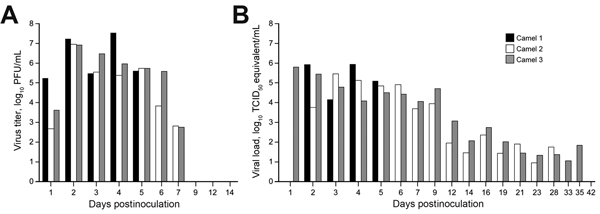
Figure 2. Virus shedding from the upper respiratory tract in dromedary camels inoculated Middle East respiratory syndrome coronavirus (MERS-CoV). Shedding was determined by A) infectious titers by plaque assay and B) viral load by quantitative real-time PCR. We extrapolated 50% tissue culture infective dose (TCID50) equivalents from standard curves generated by 10-fold dilutions of a MERS-CoV stock (HCoV-EMC/2012) with known virus titer in parallel to each quantitative real-time PCR run.
1These senior authors contributed equally to this article.
Page created: November 18, 2014
Page updated: November 18, 2014
Page reviewed: November 18, 2014
The conclusions, findings, and opinions expressed by authors contributing to this journal do not necessarily reflect the official position of the U.S. Department of Health and Human Services, the Public Health Service, the Centers for Disease Control and Prevention, or the authors' affiliated institutions. Use of trade names is for identification only and does not imply endorsement by any of the groups named above.