Volume 20, Number 12—December 2014
Research
Replication and Shedding of MERS-CoV in Upper Respiratory Tract of Inoculated Dromedary Camels
Danielle R. Adney, Neeltje van Doremalen, Vienna R. Brown, Trenton Bushmaker, Dana Scott, Emmie de Wit, Richard A. Bowen1 , and Vincent J. Munster1
Figure 4
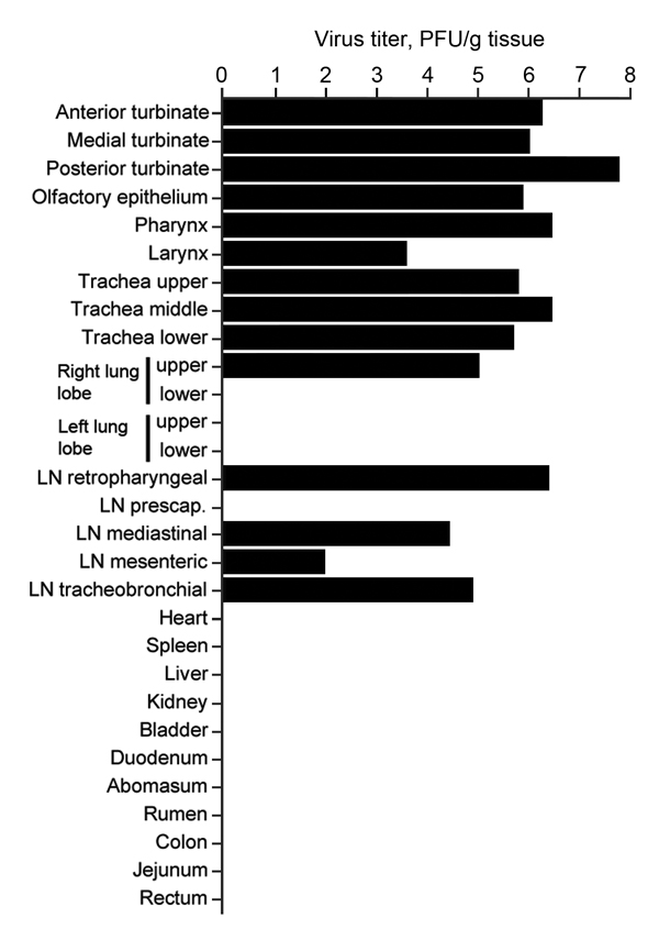
Figure 4. Virus titers in tissues collected from dromedary camel 1 inoculated with Middle East respiratory syndrome coronavirus (MERS-CoV). Tissues were collected at 5 days postinoculation (dpi) for camel 1, 28 dpi for vamel 2 and 42 dpi for camel 3. Detectable infectious virus in the collected tissues was found only in camel 1. Nasal turbinates were sampled in 3 different sections: anterior, medial, and posterior. Infectious titers were determined by plaque assay. LN, lymph node.
1These senior authors contributed equally to this article.
Page created: November 18, 2014
Page updated: November 18, 2014
Page reviewed: November 18, 2014
The conclusions, findings, and opinions expressed by authors contributing to this journal do not necessarily reflect the official position of the U.S. Department of Health and Human Services, the Public Health Service, the Centers for Disease Control and Prevention, or the authors' affiliated institutions. Use of trade names is for identification only and does not imply endorsement by any of the groups named above.