Volume 21, Number 10—October 2015
Dispatch
Detection of Mixed Infections with Plasmodium spp. by PCR, India, 2014
Cite This Article
Citation for Media
Abstract
In 8 malaria-endemic states in India, mixed Plasmodium spp. infections were detected by PCR in 17.4% (265/1,521) of blood samples that microscopy had shown to contain only P. falciparum. The quality of microscopy must be improved because use of PCR for detection of malaria parasites is limited in rural areas.
Five Plasmodium species (P. falciparum, P. vivax, P. malariae, P. ovale, and P. knowlesi) cause human malaria. Malaria is not uniformly distributed in India; 8 of the 35 states and union territories contain most malaria cases (1). Infections with P. falciparum and P. vivax occur at approximately equal frequencies (2–4). This finding increases the possibility of mixed infections, as reported in other countries, such as Peru (5), Papua New Guinea (6), Brazil (7), and Ethiopia (8).
In India, malaria control usually involves vector control with indoor residual spraying of insecticides and insecticide-treated bed nets, and chemotherapy with artemisinin-based combination therapy. Malaria diagnosis is based mainly on microscopic detection of parasites in peripheral blood smears from symptomatic persons. In addition, bivalent, rapid diagnostic tests (RDTs) are useful detection tools (9) but cannot differentiate P. falciparum moninfections from co-infections with other Plasmodium species (2,3). Moreover, genetic polymorphisms in diagnostic antigens limits detection by monoclonal antibodies. Misdiagnoses might also arise from gene deletions that prevent expression of proteins by the parasite (10). We report that a high proportion of mixed infections with 4 Plasmodium species detected by PCR in 8 states of India to which malaria is highly endemic were not detected by bivalent RDTs and microscopy.
This study was approved by the Institutional review board of the National Institute for Research in Tribal Health (Jabalpur, India). Written informed consent was obtained from all participants or parents of children, according to Indian Council of Medical Research guidelines.
Figure 1
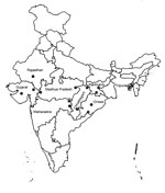
Figure 1. Fifteen community health centers in 8 states in India to which malaria is endemic. 1, Udaipur; 2, Dahod; 3, Valsad; 4, Jhabua; 5, Annupur; 6, Gondia; 7, Gadchiroli; 8, Jagdalpur; 9,...
The study was conducted in 2 community health centers (CHCs), 1 in an area that had a high level of malaria endemicity and 1 that had a low level of malaria endemicity, in each of 8 states in India: Orissa, Chhattisgarh, Jharkhand, Maharashtra, Madhya Pradesh, Tripura, Gujarat, and Rajasthan (Figure 1; Table 1). Selected CHCs were located in different regions, and forest areas in these regions ranged from 13% in Jhabua (Madhya Pradesh) to 81% in Tripura. Elevation above sea level ranged from 13 m in Valsad (Gujarat) to 870 m in Koraput (Orissa). Inhabitants of most study areas were ethnic tribes (39%–87%). All areas had received 2 rounds of indoor residual spray (DDT/synthetic pyrethroid) as a vector control measure.
Blood samples were collected from persons with suspected malaria during July–December 2014 at malaria clinics in CHC hospitals at 15 sites. Microscopy and RDTs (Bioline Ag Malaria Pf/Pv Test; Standard Diagnostics Inc., Gyeonggi-do, South Korea) were performed at outpatient department clinics of CHCs, and molecular diagnosis (PCR and sequencing) was performed at the molecular parasitology laboratory at the National Institute for Research in Tribal Health.
For microscopy, thick and thin blood smears were prepared from finger prick blood samples, which were air-dried, fixed in methanol, and stained with Giemsa. A total of 100 thick blood smear fields were examined by using an oil immersion lens at 100× magnification before a sample was considered negative. Malaria parasite density was determined from thick blood smears by counting the number of parasites against 200 leukocytes (11). Microscopy was also performed on samples that had negative results by RDT. Blood smears were cross-checked by a senior laboratory technician. RDT was performed according to manufacturer’s instructions (9) and was repeated for samples in which discordant results were obtained (e.g., microscopy positive, RDT negative).
Genomic DNA was isolated from samples that microscopy showed to contain only P. falciparum by using the QIAamp DNA Blood Mini Kit (QIAGEN, Hilden, Germany). Species-specific nested PCRs that targeted the 18S rRNA gene were used to detect 4 malaria parasite species (P. falciparum, P. vivax, P. ovale, and P. malariae) (12). P. knowlesi was detected by using a set of primers specific for the 18S rRNA gene (13), and differentiation of 2 subspecies of P. ovale (P. curtisi and P. wallikeri) was performed as described (4). PCR primers and conditions are shown in Table 2. An independent research assistant, who was unaware of microscopy or RDTs results, performed PCR on coded samples.
Figure 2
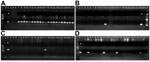
Figure 2. Identification of Plasmodium spp. by nested PCR at 15 community health centers in 8 states in India to which malaria is endemic. A) Plasmodium falciparum (205-bp fragment). Lane 1, molecular mass...
Of 1,521 samples determined by microscopy to be P. falciparum moninfections, PCR confirmed results for 1,256 (83%). However, PCR showed mixed infections with P. falciparum and P. vivax in 239 (16%) samples; P. falciparum and P. malariae in 19 (1%) samples; P. falciparum and P. ovale in 6 (0.4%) samples; and P. falciparum, P. malariae, and P. ovale in 1 (0.1%) sample (Table 1). Microscopy could not identify these mixed infections (17.4% [265/1,521]). PCR amplification of DNA from 4 Plasmodium species is shown in Figure 2.
Secondary microscopic analysis of blood smears by a second technician showed that only 22/239 (9.2%) samples contained mixed infections with P. falciparum and P. vivax. PCR analysis showed that the highest prevalence of mixed infections with P. falciparum and P. vivax was in Jharkhand (25.5%, 55/216), followed by Madhya Pradesh (20.8%, 47/226), Rajasthan (18.6%, 26/140), Orissa (15%, 40/267), and Tripura (15%, 19/127), and Chhattisgarh (10.7%, 23/214). The lowest prevalences were in Maharashtra (9%, 21/234) and Gujarat (8.2%, 8/97).
Mixed infections with P. falciparum and P. malariae were found in all 8 states, although in small numbers. Mixed infections with P. falciparum and P. ovale were found in only 4 states, particularly at CHCs in areas to which malaria was highly endemic. Of 7 mixed infections that contained P. ovale, 5 contained P. ovale curtisi and 2 contained P. ovale wallikeri. P. knowlesi was not found in any state.
This study was conducted 8 states in India that contain 80% of malaria cases (85% of which are caused by P. falciparum) and 70% of deaths caused by malaria in the entire country (1). Misdiagnosis by microscopy occurs because in mixed infections there is a tendency of 1 parasite to predominate and microscopy usually does not detect low numbers of other parasites (6). Thus, rare malaria parasites and mixed infections are underestimated through routine microscopy and RDTs (2–6), and misidentification of malaria parasites could prolong parasite clearance time and lead to anemia and drug resistance (14). A high proportion of mixed infections with P. vivax and P. falciparum have been reported in India (15). However, in that study, Gupta et al. did not look for P. malariae or P. ovale and their sample size was small (180 persons).
Our study had some limitations. Monoinfections or mixed infections were not verified by PCR if parasitemia levels were too low to be detected by microscopy or RDTs. Thus, mixed infections with parasitemia levels below the limit of detection of microscopy or RDTs would not have been detected. Detailed studies in different ecosystems during different transmission seasons and large sample sizes are required for a more accurate picture of mixed infections with common and uncommon parasite species, clinical epidemiology, adverse effects, relapse, and recrudescence.
Our results highlight the role of mixed infections, particularly those with P. vivax, P. malariae, and P. ovale, which are not detected accurately by microscopy or RDTs. Although P. vivax and P. ovale are responsible for relapses (4), P. malariae is sustained at low rates among sparse and mobile human populations for decades, thus facilitating transmission by mosquitoes (2). Our results also emphasize a major concern in the diagnosis of malaria by microscopy or RDTs and has serious repercussions for malaria epidemiology and subsequent control. These findings indicate the need to improve quality of microscopy and RDTs because PCR techniques are expensive. Until PCR becomes much less expensive and more available as a point-of-care test for field testing, its use will be limited for malaria detection.
Mr. Krishna is a senior research fellow at the National Institute for Research in Tribal Health, Jabalpur, India. His research interest is molecular characterization of malaria parasites.
Acknowledgments
We thank all study participants and their relatives for providing informed consent and staff of 15 CHC hospitals, particularly medical officers, for their help and support.
This study was supported by the Indian Council of Medical Research, New Delhi, India.
N.S. conceived the study; N.S., P.K.B., and MPS designed the study protocol; S.K., H.S.C., A.A., R.K., and P.P.S. conducted sample collection and molecular experiments; S.K., H.S.C., and P.K.B. analyzed sequencing data; N.S., M.P.S., and P.K.B., analyzed and interpreted data; and N.S., P.K.B., and M.P.S. drafted the manuscript. All authors read and approved the final manuscript.
References
- National Vector Borne Disease Control Programme. Malaria situation in India, 2014 [cited 2015 Apr 16]. http://nvbdcp.gov.in/Doc/mal_situation_Feb2015.pdf
- Bharti PK, Chand SK, Singh MP, Mishra S, Shukla MM, Singh R, Emergence of a new focus of Plasmodium malariae in forest villages of district Balaghat, central India: implications for the diagnosis of malaria and its control. Trop Med Int Health. 2013;18:12–7 . DOIPubMedGoogle Scholar
- Singh R, Jain V, Singh PP, Bharti PK, Thomas T, Basak S, First report of detection and molecular confirmation of Plasmodium ovale from severe malaria cases in central India. Trop Med Int Health. 2013;18:1416–20. DOIPubMedGoogle Scholar
- Chaturvedi N, Bhandari S, Bharti PK, Basak SK, Singh MP, Singh N. Sympatric distribution of Plasmodium ovale curtisi and P. ovale wallikeri in India: implication for the diagnosis of malaria and its control. Trans R Soc Trop Med Hyg. 2015;109:352–4. DOIPubMedGoogle Scholar
- Bharti AR, Patra KP, Chuquiyauri R, Kosek M, Gilman RH, Llanos-Cuentas A, Polymerase chain reaction detection of Plasmodium vivax and Plasmodium falciparum DNA from stored serum samples: implications for retrospective diagnosis of malaria. Am J Trop Med Hyg. 2007;77:444–6 .PubMedGoogle Scholar
- Genton B, D’Acremont V, Rare L, Baea K, Reeder JC, Alpers MP, Plasmodium vivax and mixed infections are associated with severe malaria in children: a prospective cohort study from Papua New Guinea. PLoS Med. 2008;5:e127 . DOIPubMedGoogle Scholar
- Lorenzetti A, Fornazari PA, Bonini-Domingos AC, de Souza Rodrigues Penhalbel R, Fugikaha E, Bonini-Domingos CR, Mixed Plasmodium falciparum infections and its clinical implications in four areas of the Brazilian Amazon region. Acta Trop. 2008;107:8–12. DOIPubMedGoogle Scholar
- Mekonnen SK, Aseffa A, Berhe N, Teklehaymanot T, Clouse RM, Gebru T, Return of chloroquine-sensitive Plasmodium falciparum parasites and emergence of chloroquine-resistant Plasmodium vivax in Ethiopia. Malar J. 2014;13:244. DOIPubMedGoogle Scholar
- Singh N, Bharti PK, Singh MP, Mishra S, Shukla MM, Sharma RK, Comparative evaluation of bivalent malaria rapid diagnostic tests versus traditional methods in field with special reference to heat stability testing in central India. PLoS ONE. 2013;8:e58080. DOIPubMedGoogle Scholar
- Wurtz N, Fall B, Bui K, Pascual A, Fall M, Camara C, Pfhrp2 and pfhrp3 polymorphisms in Plasmodium falciparum isolates from Dakar, Senegal: impact on rapid malaria diagnostic tests. Malar J. 2013;12:34. DOIPubMedGoogle Scholar
- World Health Organization. Parasitological confirmation of malaria diagnosis, 2009 [cited 2015 Jul 9]. http://whqlibdoc.who.int/publications/2010/9789241599412_eng.pdf
- Snounou G, Viriyakosol S, Jarra W, Thaithong S, Brown KN. Identification of the four human malaria parasite species in field samples by the polymerase chain reaction and detection of a high prevalence of mixed infections. Mol Biochem Parasitol. 1993;58:283–92 and. DOIPubMedGoogle Scholar
- Lucchi NW, Poorak M, Oberstaller J, DeBarry J, Srinivasamoorthy G, Goldman I, A new single-step PCR assay for the detection of the zoonotic malaria parasite Plasmodium knowlesi. PLoS ONE. 2012;7:e31848. DOIPubMedGoogle Scholar
- de Roode JC, Culleton R, Bell AS, Read AF. Competitive release of drug resistance following drug treatment of mixed Plasmodium chabaudi infections. Malar J. 2004;3:33. DOIPubMedGoogle Scholar
- Gupta B, Gupta P, Sharma A, Singh V, Dash AP, Das A. High proportion of mixed-species Plasmodium infections in India revealed by PCR diagnostic assay. Trop Med Int Health. 2010;15:819–24. DOIPubMedGoogle Scholar
Figures
Tables
Cite This ArticleTable of Contents – Volume 21, Number 10—October 2015
| EID Search Options |
|---|
|
|
|
|
|
|
Please use the form below to submit correspondence to the authors or contact them at the following address:
Neeru Singh, Centre for Tribals, National Institute for Research in Tribal Health, NIRTH Campus, Nagpur Rd, Garha, Jabalpur 482003, Madhya Pradesh, India
Top